The venous system of the lower limbs plays a crucial role in returning deoxygenated blood from the legs and feet back to the heart, ensuring efficient circulation and preventing issues like swelling or clots. This network includes both deep and superficial veins that work together to overcome gravity through muscle contractions and one-way valves. Understanding these structures is essential for grasping how blood flow supports overall mobility and health in the lower body.
Detailed Anatomy of Labeled Veins
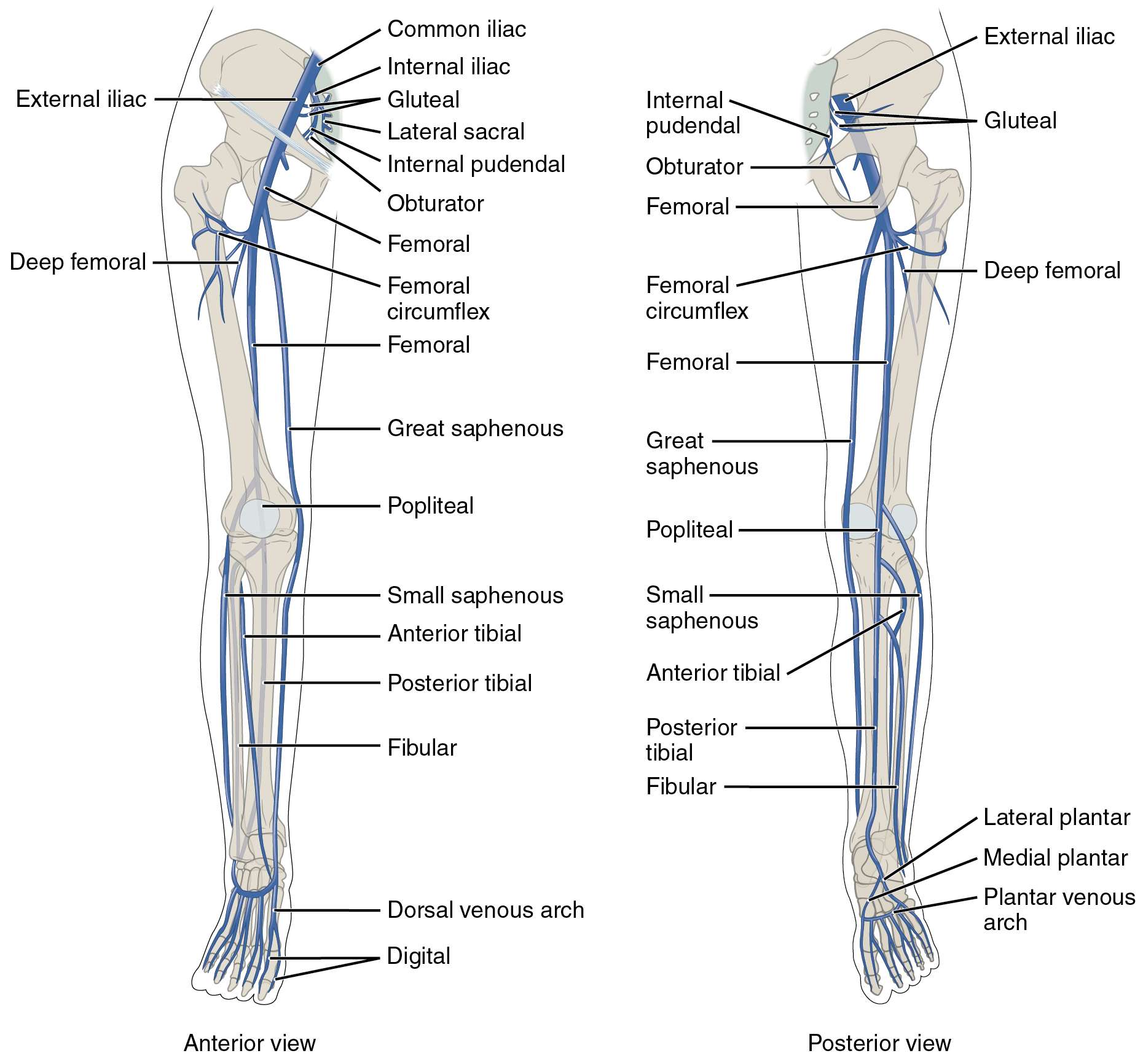
The following sections break down each labeled vein from the anterior and posterior views, highlighting their paths and contributions to venous drainage.
Common iliac: The common iliac vein forms from the union of the external and internal iliac veins at the pelvic brim, serving as a major conduit for blood returning from the lower limbs and pelvis. It ascends to join its counterpart from the opposite side, forming the inferior vena cava that leads directly to the heart.
External iliac: The external iliac vein runs alongside its arterial counterpart, collecting blood from the lower limb and continuing from the femoral vein at the inguinal ligament. This vessel is vital for draining the majority of venous blood from the leg, ensuring smooth transition into the pelvic region.
Internal iliac: The internal iliac vein drains blood from the pelvic organs, gluteal region, and parts of the thigh, merging with the external iliac to form the common iliac. Its tributaries include veins from the bladder, rectum, and reproductive organs, making it essential for pelvic venous return.
Gluteal: The gluteal veins, including superior and inferior branches, drain the gluteal muscles and surrounding tissues, accompanying the gluteal arteries. They contribute to the internal iliac vein, supporting blood flow from the buttocks area critical for posture and movement.
Lateral sacral: The lateral sacral veins collect blood from the sacral region and vertebral column, draining into the internal iliac vein. These veins help maintain circulation in the lower spine and pelvis, preventing stagnation in areas prone to pressure.
Internal pudendal: The internal pudendal vein drains the perineal region, including external genitalia and anal area, following the path of the pudendal artery. It joins the internal iliac vein, playing a key role in venous return from sensitive pelvic floor structures.
Obturator: The obturator vein drains the adductor muscles of the thigh and hip joint, passing through the obturator canal to enter the internal iliac vein. This vein ensures proper drainage from the medial thigh, supporting hip mobility and stability.
Femoral: The femoral vein is a major deep vein that continues from the popliteal vein, running through the thigh to become the external iliac at the inguinal ligament. It receives tributaries from both deep and superficial systems, making it central to lower limb venous return and vulnerable to conditions like deep vein thrombosis.
Deep femoral: The deep femoral vein, also known as the profunda femoris, parallels the deep femoral artery and drains the deeper muscles of the thigh. It joins the femoral vein, providing an important pathway for blood from the quadriceps and hamstrings.
Femoral circumflex: The femoral circumflex veins, including medial and lateral branches, drain the upper thigh and hip region, accompanying the circumflex arteries. They typically empty into the femoral or deep femoral veins, aiding in circulation around the hip joint.
Great saphenous: The great saphenous vein is the longest superficial vein in the body, starting from the dorsal venous arch of the foot and ascending medially to join the femoral vein at the saphenous opening. It drains the medial and anterior aspects of the leg and thigh, often used in surgical procedures like bypass grafts due to its accessibility.
Popliteal: The popliteal vein forms behind the knee from the union of anterior and posterior tibial veins, continuing as the femoral vein. It drains the knee joint and calf muscles, with valves that prevent backflow during standing or walking.
Small saphenous: The small saphenous vein is a superficial vessel that runs along the posterior leg from the lateral foot to the popliteal fossa, where it joins the popliteal vein. It primarily drains the lateral and posterior aspects of the lower leg, complementing the great saphenous in superficial drainage.
Anterior tibial: The anterior tibial vein drains the anterior compartment of the leg, including the dorsiflexor muscles, and ascends to unite with the posterior tibial to form the popliteal. Paired with its artery, it ensures blood return from the shin area essential for foot movement.
Posterior tibial: The posterior tibial vein runs deep in the calf, draining the plantar aspect of the foot and posterior leg muscles, joining the anterior tibial at the popliteal. It receives perforating veins from superficial systems, playing a pivotal role in pumping blood upward via calf muscle action.
Fibular: The fibular vein, also called peroneal, accompanies the fibular artery in the lateral calf, draining deep tissues and merging with the posterior tibial. This vein supports circulation in the lateral leg compartment, aiding in balance and peroneal muscle function.
Dorsal venous arch: The dorsal venous arch is a network of veins on the dorsum of the foot that collects blood from the toes and foot surface. It gives rise to the great and small saphenous veins, serving as the starting point for superficial venous return in the lower limb.
Digital: The digital veins drain the toes, forming arcs on both dorsal and plantar sides of the foot. They feed into the venous arches, ensuring efficient clearance of deoxygenated blood from the distal extremities.
Lateral plantar: The lateral plantar vein drains the lateral sole of the foot, accompanying the lateral plantar artery and joining the plantar venous arch. It supports weight-bearing areas, contributing to overall foot venous drainage.
Medial plantar: The medial plantar vein collects blood from the medial sole, running with the medial plantar artery to enter the plantar venous arch. This vein is crucial for maintaining circulation in the arch of the foot during locomotion.
Plantar venous arch: The plantar venous arch forms a network under the foot, receiving digital and plantar veins before continuing as the posterior tibial. It acts as a reservoir for blood during standing, with muscle pumps aiding return flow.
Physiological Role of Lower Limb Veins
The veins in the lower limbs are designed to handle the challenges of gravity, relying on muscular contractions and valves to propel blood upward. This system integrates deep and superficial pathways for redundant and efficient drainage.
- Deep veins like the femoral and popliteal carry the bulk of venous blood, protected within muscle compartments to benefit from the skeletal muscle pump during activities such as walking.
- Superficial veins, including the great and small saphenous, provide an alternative route and connect to deep veins via perforators, allowing for heat regulation and additional capacity during exercise.
- Valves within these veins prevent reflux, maintaining unidirectional flow toward the heart and reducing the risk of venous insufficiency.
- Blood from the feet ascends through arches and tibial veins, merging at key junctions to enter the thigh and pelvis efficiently.
Clinical Significance and Common Issues
Understanding venous anatomy helps in diagnosing and treating lower limb conditions, from varicose veins to more serious thromboses. Preventive measures focus on promoting healthy circulation through lifestyle choices.
- Varicose veins often affect the great saphenous, leading to bulging and discomfort due to valve failure and increased pressure.
- Deep vein thrombosis typically involves the femoral or popliteal veins, where clots can form from immobility, potentially leading to pulmonary embolism if dislodged.
- Surgical interventions, such as vein stripping or ablation, target problematic superficial veins while preserving deep pathways.
- Compression therapy and exercise enhance venous return, mimicking the natural muscle pump to alleviate symptoms like edema.
Integration with Overall Circulatory System
The lower limb veins connect seamlessly with the pelvic and abdominal systems, ensuring whole-body homeostasis. This integration supports nutrient delivery and waste removal across tissues.
- Upon reaching the common iliac, blood mixes with pelvic drainage before entering the inferior vena cava, which transports it to the right atrium.
- Hormonal influences, such as those from the adrenal glands releasing cortisol, can affect vascular tone and fluid balance in these veins.
- During pregnancy or prolonged standing, increased pressure on iliac veins may cause temporary swelling, highlighting the need for movement to aid flow.
- Lymphatic vessels parallel many of these veins, working in tandem to manage interstitial fluid and immune responses in the lower body.
In summary, the major veins of the lower limbs form a sophisticated network that not only returns blood against gravity but also adapts to daily demands of movement and posture. Appreciating this anatomy fosters better awareness of circulatory health, encouraging practices that sustain efficient venous function throughout life.

