Discover the intricate network of veins in the upper limb with this detailed guide, featuring an anterior view that highlights the major veins draining this region. This article provides a comprehensive look at the anatomy, function, and clinical relevance of these vascular structures, making it an essential resource for understanding human physiology.
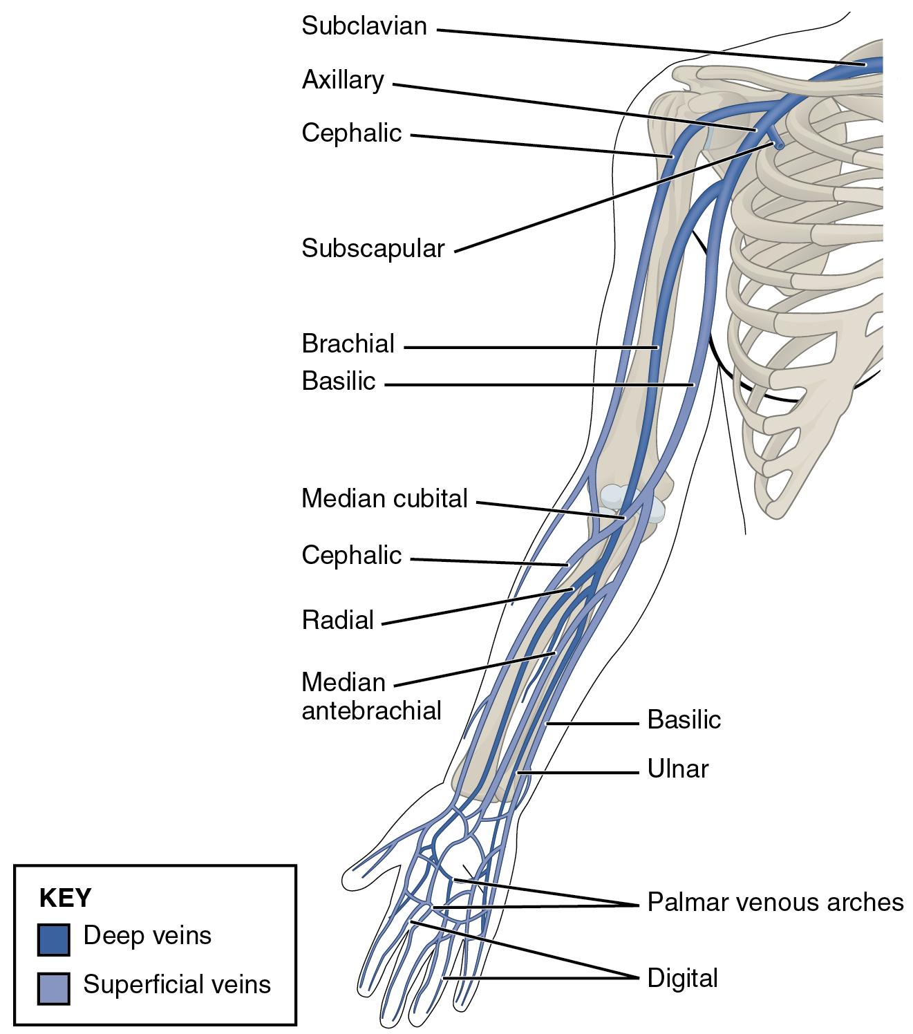
Subclavian The subclavian vein is a major vessel that begins at the lateral border of the first rib, receiving blood from the axillary vein. It plays a critical role in returning deoxygenated blood from the upper extremity to the heart.
Axillary The axillary vein runs through the axilla, formed by the union of the basilic and brachial veins, and continues as the subclavian vein. It is essential for draining blood from the upper arm and shoulder region.
Cephalic The cephalic vein originates from the dorsal venous network of the hand and travels along the lateral aspect of the arm, eventually joining the axillary vein. It is commonly used for venipuncture due to its superficial location and accessibility.
Subscapular The subscapular vein drains the region around the scapula, contributing to the axillary vein system. It is a smaller tributary that supports venous return from the shoulder area.
Brachial The brachial vein accompanies the brachial artery along the upper arm, typically existing as a pair of veins. It collects blood from the deep tissues of the arm and merges with the basilic vein to form the axillary vein.
Basilic The basilic vein runs along the medial side of the forearm and arm, joining the brachial vein to form the axillary vein. It is another key site for medical procedures like intravenous access.
Median cubital The median cubital vein connects the cephalic and basilic veins in the cubital fossa, making it a prominent site for blood draws. Its superficial position and large size enhance its clinical utility.
Radial The radial vein follows the radial artery along the lateral forearm, draining blood from the lateral hand and wrist. It is part of the deep venous system and merges with the ulnar vein to form the brachial vein.
Median antebrachial The median antebrachial vein drains the forearm and often connects the cephalic and basilic veins. It serves as an important superficial vein for venous return in the lower arm.
Ulnar The ulnar vein parallels the ulnar artery on the medial forearm, collecting blood from the medial hand and wrist. Like the radial vein, it contributes to the deep venous system.
Palmar venous arches The palmar venous arches are networks of veins in the palm that receive blood from the digital veins and drain into the radial and ulnar veins. They ensure efficient venous drainage from the hand.
Digital The digital veins drain blood from the fingers, feeding into the palmar venous arches. They are crucial for maintaining circulation in the digits.
Anatomical Overview of Upper Limb Veins
Understanding the venous system of the upper limb begins with recognizing its dual structure. The veins are categorized into deep and superficial systems, each with distinct roles.
- The deep veins, such as the radial, ulnar, and brachial veins, run alongside arteries and drain the deeper tissues of the arm.
- Superficial veins, including the cephalic, basilic, and median cubital, lie closer to the skin and are vital for clinical procedures.
- These systems work together to return deoxygenated blood to the heart, supported by valves that prevent backflow.
- The transition from superficial to deep veins, such as the axillary and subclavian, ensures efficient circulation.
Clinical Significance and Functions
The venous anatomy of the upper limb has significant medical implications. Each vein serves a purpose beyond mere drainage.
- The cephalic and basilic veins are frequently accessed for intravenous therapy or blood sampling due to their size and location.
- Deep vein thrombosis (DVT) can affect veins like the brachial or axillary, potentially leading to serious complications if untreated.
- The median cubital vein’s prominence makes it a primary choice for phlebotomy, reducing patient discomfort.
- Proper understanding of these veins aids in diagnosing circulatory issues and planning surgical interventions.
Detailed Anatomy and Physical Characteristics
The physical structure of upper limb veins varies by type and location. This section explores their unique features.
- Deep veins, such as the radial and ulnar, are typically paired and encased in connective tissue with arteries, providing structural support.
- Superficial veins like the subclavian and axillary are larger and more elastic, accommodating varying blood volumes.
- The digital veins are smaller and more numerous, ensuring thorough drainage from the fingers.
- Valves within these veins, particularly in the cephalic and basilic, regulate blood flow against gravity.
Practical Applications in Medicine
Knowledge of upper limb veins is essential for various medical practices. This section highlights their use.
- The subscapular and median antebrachial veins are considered in procedures involving the shoulder or forearm.
- Catheter insertion often targets the axillary or subclavian veins for central venous access.
- Understanding venous patterns helps in managing trauma or injuries to the palmar venous arches.
- Regular monitoring of these veins can prevent complications like edema or varicose veins.
Conclusion
The veins of the upper limb form a complex yet fascinating network that supports circulation and offers critical access points for medical intervention. From the digital veins in the fingers to the subclavian vein near the heart, each plays a vital role in maintaining health. This guide provides a solid foundation for exploring their anatomy and clinical applications, encouraging further study to enhance practical skills in healthcare.

