The lower limb’s arterial system is a vital network that ensures oxygen-rich blood reaches the legs and feet, supporting movement and overall health. This anterior view image highlights the major arteries, tracing their path from the pelvis down to the toes, offering a clear understanding of how circulation sustains this critical region of the body.
Exploring the Key Arteries in the Anterior View
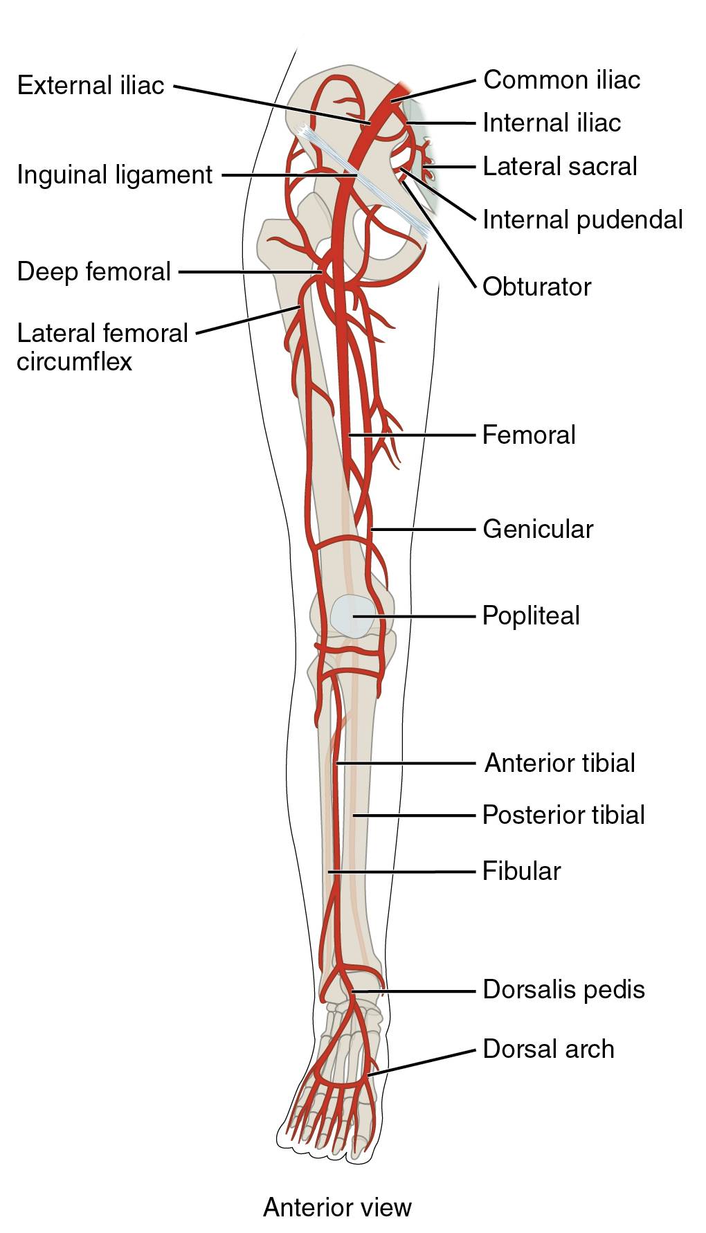
The labeled arteries in this image provide a detailed map of blood supply to the lower limb from an anterior perspective. Each vessel contributes uniquely to the vascular network, ensuring efficient nutrient delivery.
External iliac The external iliac artery branches from the common iliac, transitioning into the femoral artery below the inguinal ligament. It serves as the primary conduit for blood flow to the lower limb, supporting pelvic and thigh circulation.
Common iliac The common iliac artery arises from the abdominal aorta, splitting into internal and external iliac arteries. It delivers blood to the pelvic region and lower extremity, forming a crucial junction in the vascular system.
Internal iliac The internal iliac artery branches from the common iliac, supplying the pelvic organs and medial thigh muscles. It supports reproductive, urinary, and muscular functions through its extensive branching.
Lateral sacral The lateral sacral arteries stem from the internal iliac, providing blood to the sacral region and spinal nerves. They enhance lower back stability and support sacral nerve function.
Internal pudendal The internal pudendal artery, a branch of the internal iliac, supplies the external genitalia and perineum. It ensures adequate perfusion for reproductive and anal structures, aiding their physiological roles.
Obturator The obturator artery arises from the internal iliac, passing through the obturator foramen to supply the medial thigh. It nourishes the adductor muscles and contributes to hip joint stability.
Inguinal ligament The inguinal ligament acts as a landmark where the external iliac artery becomes the femoral artery. It marks the transition from the pelvic to the thigh vascular territory.
Deep femoral The deep femoral artery branches from the femoral, supplying the deep thigh muscles and hip joint. It provides a robust collateral circulation, enhancing blood flow to the posterior thigh.
Lateral femoral circumflex The lateral femoral circumflex artery branches from the deep femoral, encircling the femur to supply lateral thigh muscles. It supports hip abduction and thigh movement.
Femoral The femoral artery, a continuation of the external iliac, runs down the anterior thigh, supplying the quadriceps and other muscles. It is a key site for pulse palpation in the groin.
Genicular The genicular arteries branch from the femoral and popliteal, forming a network around the knee. They ensure blood supply to the knee joint, supporting mobility and healing.
Popliteal The popliteal artery continues from the femoral behind the knee, supplying the knee and calf muscles. It bifurcates into the anterior and posterior tibial arteries, critical for lower leg flow.
Anterior tibial The anterior tibial artery branches from the popliteal, running along the shin to supply anterior leg muscles. It continues as the dorsalis pedis, feeding the foot’s dorsal surface.
Posterior tibial The posterior tibial artery, another popliteal branch, runs along the calf, supplying posterior muscles and the foot sole. It contributes to the plantar arch, ensuring foot perfusion.
Fibular The fibular artery branches from the posterior tibial, supplying the lateral leg and ankle. It provides collateral circulation, supporting the fibular muscles.
Dorsalis pedis The dorsalis pedis artery extends from the anterior tibial, running across the foot’s top. It supplies the dorsal foot and contributes to the dorsal arch.
Dorsal arch The dorsal arch is formed by the dorsalis pedis and other arteries, supplying the toes and dorsal foot. It ensures comprehensive blood distribution to the foot’s upper region.
The Pathway of Lower Limb Arteries
Blood flow to the lower limb originates from the abdominal aorta, following a structured route to the extremities. This pathway adapts to the leg’s functional demands.
- The aorta bifurcates into the common iliac arteries, initiating lower limb circulation.
- The external iliac becomes the femoral artery, transitioning at the inguinal ligament.
- The popliteal artery splits into anterior and posterior tibial branches behind the knee.
- Distal arteries like the dorsalis pedis ensure foot and toe perfusion.
Clinical Relevance of Lower Limb Arteries
Understanding these arteries aids in diagnosing vascular conditions. Their anatomical positions guide clinical assessments.
- The femoral artery is used for arterial access during catheterization and pulse checks.
- The dorsalis pedis pulse assesses foot perfusion, crucial in peripheral artery disease.
- Genicular arteries support knee surgeries by providing collateral flow.
- Ultrasound evaluates flow in the popliteal and tibial arteries for blockages.
Physiological Roles in Lower Limb Function
These arteries adapt to physical activity, ensuring oxygen delivery. Their responsiveness enhances mobility and endurance.
- The femoral and deep femoral arteries dilate during exercise, boosting thigh muscle performance.
- The dorsal arch regulates blood flow to support standing and walking.
- Hormonal influences, like adrenaline, modulate vascular tone in the lower limb.
- Anastomoses around the knee protect against ischemic events.
In conclusion, the anterior view of the lower limb’s major arteries reveals a resilient network supporting movement and stability. Mastering this anatomy enhances clinical practice and deepens appreciation for circulatory efficiency.

