Major Arteries Serving the Thorax and Upper Limb: A Comprehensive Anatomical Guide
The human upper limb relies on a complex network of arteries to deliver oxygen-rich blood from the heart to the muscles, bones, and tissues of the arm and hand. This intricate vascular system begins at the subclavian artery and branches extensively to ensure efficient circulation, supporting everything from fine motor skills to overall limb function.
Understanding the Key Arteries in the Upper Limb
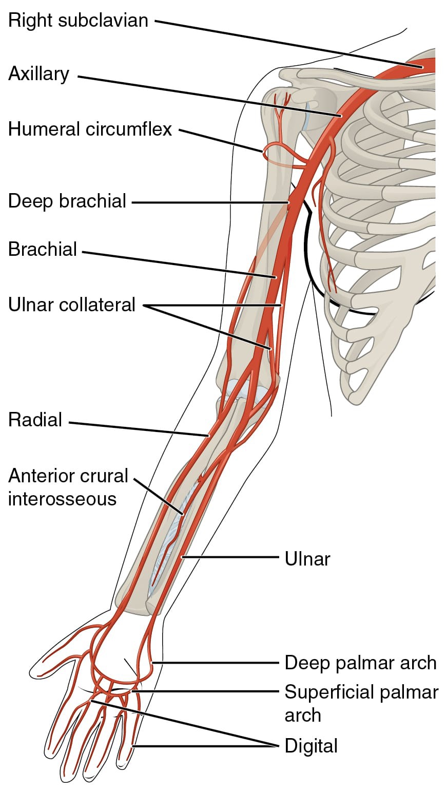
The arteries depicted in this image form the primary pathway for blood supply to the thorax and upper limb, originating from the aortic arch and extending distally. Each labeled structure plays a vital role in maintaining adequate perfusion, with variations in branching patterns that can impact clinical assessments.
Right subclavian The right subclavian artery arises from the brachiocephalic trunk and supplies blood to the right upper limb, neck, and parts of the thorax. It transitions into the axillary artery after passing under the clavicle, ensuring continuous blood flow to distal regions.
Axillary The axillary artery continues from the subclavian artery, running through the axilla region and providing branches to the shoulder and chest wall. It is divided into three parts based on its relation to the pectoralis minor muscle, each giving off important tributaries for local tissue nourishment.
Humeral circumflex The humeral circumflex arteries, including anterior and posterior branches, encircle the surgical neck of the humerus to supply the shoulder joint and surrounding muscles. These vessels anastomose with other arteries, creating collateral pathways that protect against occlusion in the main axillary trunk.
Deep brachial The deep brachial artery, also known as the profunda brachii, branches from the brachial artery and travels along the humerus to nourish the triceps muscle and posterior arm compartment. It participates in anastomoses around the elbow, contributing to the robustness of upper limb circulation.
Brachial The brachial artery is the main continuation of the axillary artery down the arm, palpable in the antecubital fossa for blood pressure measurements. It bifurcates into the radial and ulnar arteries at the elbow, serving as a critical conduit for arterial blood to the forearm and hand.
Ulnar collateral The ulnar collateral arteries, superior and inferior, arise from the brachial artery and form anastomoses around the elbow joint with branches from the radial artery. These vessels provide alternative routes for blood flow, essential during joint flexion or in cases of proximal arterial narrowing.
Radial The radial artery descends along the lateral forearm, easily felt at the wrist for pulse checks, and contributes to the deep palmar arch in the hand. It supplies blood to the thumb and lateral fingers, playing a key role in hand dexterity and sensation.
Anterior interosseous The anterior interosseous artery branches from the common interosseous artery in the forearm, running between the radius and ulna to supply deep flexor muscles and bones. It perforates the interosseous membrane to anastomose with posterior vessels, enhancing nutrient delivery to the anterior compartment.
Ulnar The ulnar artery travels medially along the forearm, giving off branches to the flexor muscles and forming the superficial palmar arch. It is crucial for blood supply to the medial fingers and hypothenar eminence, supporting grip strength and fine movements.
Deep palmar arch The deep palmar arch is formed primarily by the radial artery and completed by the deep branch of the ulnar artery, lying beneath the flexor tendons in the palm. It gives rise to metacarpal arteries that nourish the interosseous muscles and joints of the hand.
Superficial palmar arch The superficial palmar arch is mainly contributed by the ulnar artery and often completed by branches from the radial artery, positioned just under the palmar aponeurosis. It supplies common digital arteries to the fingers, ensuring adequate perfusion for tactile functions and wound healing.
Digital The digital arteries arise from the palmar arches and run along the sides of the fingers to provide blood to the phalanges and nail beds. These small vessels are vital for fingertip sensitivity and temperature regulation, with rich anastomoses that maintain flow even under pressure.
The Origin and Pathway of Upper Limb Arteries
Blood flow to the upper limb starts from the aortic arch, branching into the subclavian arteries that extend outward. This systematic progression ensures that oxygenated blood reaches even the most distal parts efficiently.
- The subclavian artery originates differently on each side: the right from the brachiocephalic trunk and the left directly from the aorta.
- As it passes laterally, it gives off branches like the vertebral and internal thoracic arteries before becoming the axillary.
- In the axilla, the vessel is surrounded by brachial plexus nerves, highlighting the integrated neurovascular bundle.
- Distal branching patterns can vary, with anomalies occurring in about 30% of individuals, affecting surgical planning.
Branching Patterns and Anastomoses
Arterial branches in the upper limb create a network of connections that safeguard against blockages. These anastomoses allow blood to reroute, maintaining tissue viability.
- Around the shoulder, the humeral circumflex arteries link with thoracoacromial branches for collateral circulation.
- Elbow anastomoses involve ulnar collateral and radial recurrent arteries, forming a protective ring.
- In the hand, deep and superficial palmar arches interconnect, providing dual supply to digits.
- Such redundancies are crucial in conditions like atherosclerosis, where gradual narrowing prompts collateral development.
Clinical Significance of Upper Limb Arteries
Knowledge of these arteries aids in diagnosing and treating vascular issues. Pulse palpation and imaging rely on understanding their locations.
- The brachial artery is key for auscultating blood pressure using a sphygmomanometer.
- Radial and ulnar pulses assess distal perfusion, important in trauma evaluations.
- Arterial injuries from fractures can lead to compartment syndrome if not addressed promptly.
- Doppler ultrasound visualizes flow in these vessels, detecting stenoses or aneurysms.
Functions in Daily Physiology
These arteries support not just survival but also performance in physical activities. They adapt to demands like exercise by vasodilating to increase blood delivery.
- Muscles in the arm receive nutrients via branches like the deep brachial, enabling sustained contraction.
- Hand arteries ensure precise control for tasks requiring dexterity, from writing to playing instruments.
- Thermoregulation involves digital vessels constricting or dilating in response to temperature changes.
- Hormonal influences, such as adrenaline, can modulate flow through these pathways.
In summary, the major arteries of the thorax and upper limb represent a marvel of evolutionary design, balancing efficiency with resilience to ensure uninterrupted blood supply. Appreciating this anatomy enhances our understanding of human physiology and informs medical interventions.

