The velocity of blood flow through various vessels in the human body plays a crucial role in maintaining efficient circulation and nutrient delivery. This graph illustrates how blood speed changes from large arteries to tiny capillaries and back through veins, highlighting the intricate balance of anatomy and physiology that supports overall cardiovascular health.
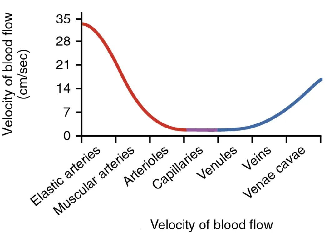
Elastic arteries These large vessels, such as the aorta, are designed to handle high-pressure blood ejected from the heart. Their elastic walls expand and recoil to smooth out pulsatile flow, ensuring a steady supply to downstream vessels.
Muscular arteries Also known as distributing arteries, these medium-sized vessels regulate blood distribution to organs through smooth muscle contraction. They maintain consistent pressure by adjusting diameter, which is vital for responding to the body’s varying demands.
Arterioles As the smallest arteries, arterioles act as resistance vessels that control blood flow into capillary beds. Their constriction or dilation fine-tunes local perfusion, directly influencing tissue oxygenation and nutrient exchange.
Capillaries These microscopic vessels form extensive networks where gas and nutrient exchange occurs between blood and tissues. With the slowest velocity, capillaries allow ample time for diffusion processes essential to cellular function.
Venules Small veins that collect blood from capillary networks, venules begin the return journey to the heart with gradually increasing flow speed. They facilitate the drainage of deoxygenated blood while allowing some exchange of substances.
Veins Medium to large conduits equipped with valves, veins transport blood back under lower pressure. Their collapsible walls and reliance on muscle pumps ensure efficient return despite gravitational challenges.
Venae cavae The largest veins, including superior and inferior types, converge blood from the body into the right atrium. They handle high-volume flow at moderate speeds, completing the systemic circuit.
The Physics Behind Blood Flow Velocity
Blood flow velocity refers to the speed at which blood travels through vessels, governed by principles like continuity and resistance. This velocity varies inversely with the total cross-sectional area of the vessels, explaining the dramatic slowdown in capillaries.
- In larger arteries, high velocity ensures rapid transport from the heart to peripheral tissues.
- The equation for flow rate, Q = A × v (where Q is flow, A is area, v is velocity), demonstrates why velocity drops as area increases in branching networks.
- Physiological factors such as viscosity and vessel length further modulate speed, impacting conditions like hypertension.
- Understanding these dynamics aids in comprehending how the body maintains homeostasis during exercise or rest.
Anatomical Variations in Blood Vessels
The structure of blood vessels is tailored to their function, with layers like tunica intima, media, and adventitia adapting accordingly. From elastic to muscular types, each segment optimizes flow characteristics for efficiency.
- Elastic arteries feature abundant elastin fibers in the media layer, allowing stretch and recoil with each heartbeat.
- Muscular arteries have thicker smooth muscle layers for vasoregulation, distributing blood based on metabolic needs.
- Arterioles possess a high muscle-to-lumen ratio, enabling precise control over resistance and pressure gradients.
- Capillaries consist of a single endothelial layer, facilitating diffusion while minimizing barriers to exchange.
- Venules and veins incorporate valves to prevent backflow, especially in lower extremities.
- The venae cavae have wide lumens to accommodate large volumes without significant resistance.
Physiological Implications of Velocity Changes
Changes in blood flow velocity influence oxygen delivery and waste removal, critical for organ function. The graph’s curve from high to low and back up reflects adaptive mechanisms in circulation.
- In arterioles, velocity reduction prepares blood for capillary exchange by increasing transit time.
- Slow flow in capillaries enhances nutrient absorption and gas diffusion, supporting cellular metabolism.
- Acceleration in venules and veins relies on skeletal muscle contraction and respiratory pumps.
- Hormonal influences, such as adrenaline, can alter vessel diameter and thus velocity.
- Disruptions in this pattern may signal issues like atherosclerosis, affecting overall perfusion.
Factors Influencing Blood Flow Dynamics
Multiple elements, including cardiac output and vascular tone, dictate blood velocity patterns. External factors like posture or hydration also play roles in maintaining optimal flow.
- Cardiac output directly correlates with initial velocity in elastic arteries.
- Vascular resistance, primarily in arterioles, inversely affects downstream speeds.
- Blood viscosity, altered by hematocrit levels, can slow flow if elevated.
- Autoregulation ensures tissues receive consistent supply despite systemic changes.
- In pathological states, such as in diabetes, microvascular changes impair capillary velocity.
Clinical Relevance and Applications
Insights from blood flow velocity graphs inform diagnostic and therapeutic strategies in medicine. Monitoring these parameters helps in managing cardiovascular conditions effectively.
- Doppler ultrasound measures velocity to detect stenoses or aneurysms in arteries.
- In critical care, understanding venous return velocity guides fluid management.
- Pharmacological interventions target vessel dilation to optimize flow in hypertension.
- Exercise physiology leverages velocity changes to enhance endurance training.
- Research into microcirculation focuses on capillary dynamics for drug delivery systems.
The velocity of blood flow encapsulates the elegance of the circulatory system’s design, ensuring life-sustaining processes occur seamlessly. By appreciating these patterns, one gains deeper insight into how the body adapts to demands, paving the way for advancements in health and wellness.

