The skeletal muscle pump is a vital mechanism that aids in returning blood to the heart, particularly in the lower extremities where gravity poses a challenge. This diagram illustrates how muscle contraction and relaxation, along with one-way valves, work together to facilitate efficient venous circulation and maintain cardiovascular health.
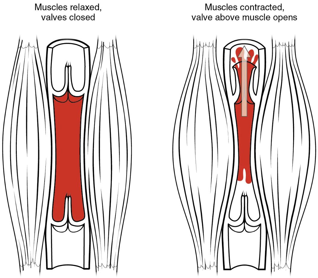
Muscles relaxed, valves closed Muscles relaxed, valves closed depicts the state when surrounding skeletal muscles are at rest, allowing venous pressure to equalize and valves to prevent backflow. This phase ensures that blood remains in place until the next muscle action, maintaining directional flow toward the heart.
Muscles contracted, valve above muscle opens Muscles contracted, valve above muscle opens shows the active phase where muscle contraction compresses the vein, increasing internal pressure and opening the valve above. This action propels blood upward toward the heart, with the valve closing to prevent reverse flow once the muscle relaxes.
Overview of the Skeletal Muscle Pump
The skeletal muscle pump plays a crucial role in venous return, especially during physical activity. Its mechanism leverages muscle movement to overcome gravitational challenges in the circulatory system.
- Muscles relaxed, valves closed allows blood to pool temporarily, preparing for the next contraction.
- Muscles contracted, valve above muscle opens drives blood upward, aided by one-way valves.
- This pump is most active in the legs, where calf muscles act like a second heart.
- Valves ensure unidirectional flow, critical for preventing venous stasis.
- The process is enhanced by rhythmic movements like walking or exercise.
Anatomical Role in Venous Circulation
The skeletal muscle pump integrates with the venous system to support blood return. Its anatomical design optimizes efficiency in the face of gravitational pull.
- Muscles relaxed, valves closed occurs when skeletal muscles, such as the gastrocnemius, are inactive, letting valves seal the vein.
- Muscles contracted, valve above muscle opens involves compression of the vein by contracting muscles, forcing blood past the upper valve.
- Veins in the lower limbs, like the popliteal and femoral veins, rely heavily on this mechanism.
- The presence of bicuspid valves prevents reflux, maintaining pressure gradients.
- This system complements the heart’s pumping action, especially in upright postures.
Mechanism of Muscle Contraction
Muscle contraction is the driving force behind the skeletal muscle pump. This action transforms mechanical movement into circulatory support.
- Muscles contracted, valve above muscle opens squeezes the vein, raising internal pressure to 20-30 mmHg.
- The force opens the valve, allowing blood to move toward lower pressure areas like the vena cava.
- Relaxation phase, muscles relaxed, valves closed, sees valves close due to backpressure, halting reverse flow.
- The calf muscle pump can increase venous return by up to 60% during exercise.
- Sympathetic nervous system activation enhances this effect during physical stress.
Importance of Valves in Blood Flow
Valves are essential for directing blood flow in the skeletal muscle pump. Their function ensures efficient circulation against gravity.
- Muscles relaxed, valves closed maintains blood position, with valves sealing due to slight backpressure.
- Muscles contracted, valve above muscle opens allows forward flow, with the valve above opening under increased pressure.
- Malfunctioning valves can lead to conditions like varicose veins or chronic venous insufficiency.
- Each valve is composed of endothelial flaps, responding to pressure changes dynamically.
- Regular valve function is vital for preventing blood pooling in dependent areas.
Clinical Significance and Applications
The skeletal muscle pump’s efficiency has implications for both health maintenance and disease prevention. Understanding its action aids in clinical assessments and interventions.
- Weak muscles relaxed, valves closed phases may indicate muscle atrophy, reducing pump effectiveness.
- Enhanced muscles contracted, valve above muscle opens activity improves circulation in sedentary individuals.
- Prolonged inactivity can impair this mechanism, increasing deep vein thrombosis risk.
- Physical therapy often targets this pump to aid recovery in post-surgical patients.
- Monitoring venous return helps assess cardiovascular fitness and guide exercise prescriptions.
In conclusion, the skeletal muscle pump diagram highlights a sophisticated interplay between muscles relaxed, valves closed and muscles contracted, valve above muscle opens to ensure effective venous return. This mechanism underscores the body’s ability to adapt circulation to varying conditions, supporting overall cardiovascular health. By appreciating this process, one can better understand its role in daily activity and its potential impact on long-term wellness.

