The elastic artery, a key component of the circulatory system, serves as a conduit for oxygenated blood from the heart, adapting to the high-pressure demands of each heartbeat. This image focuses on the tunica intima, tunica media, tunica adventitia, and elastic lamellae, highlighting the specialized features that enable these large vessels, such as the aorta, to maintain consistent blood flow.
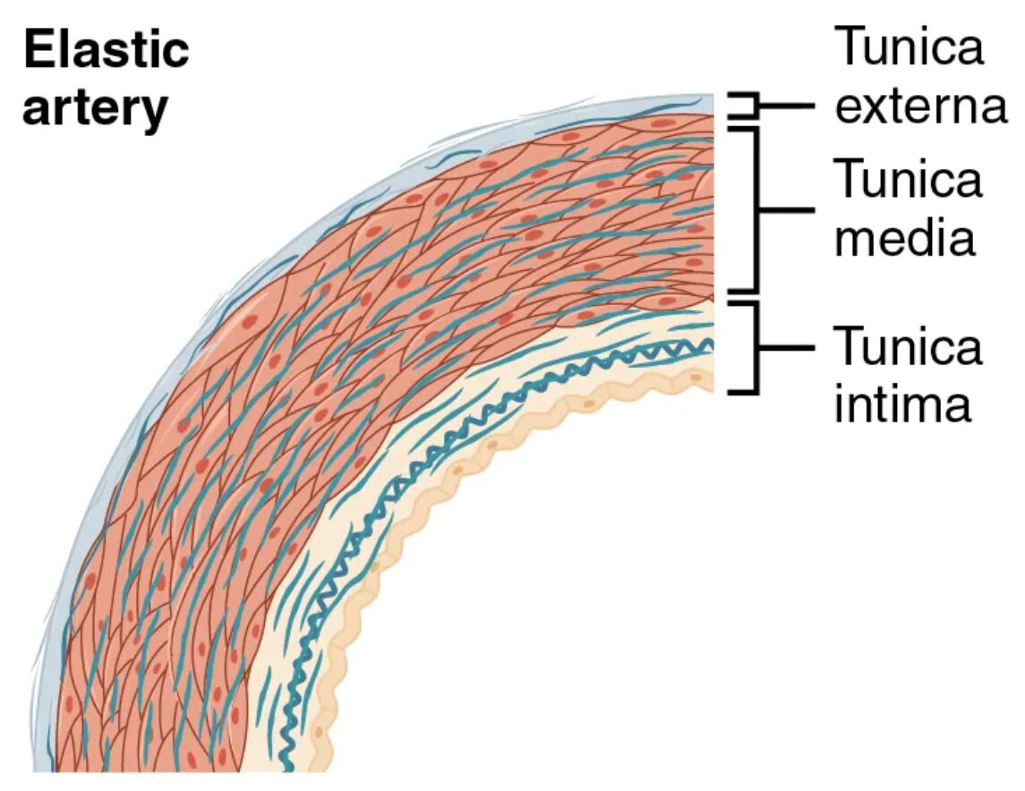
Tunica intima Tunica intima is the innermost layer, featuring a single layer of endothelial cells that line the lumen and reduce friction during blood flow. This layer includes a subendothelial layer and a thick internal elastic lamina, which enhances the artery’s ability to stretch and recoil with each cardiac cycle.
Tunica media Tunica media forms the thick middle layer, dominated by numerous elastic lamellae interspersed with smooth muscle cells. This structure absorbs the high systolic pressure and facilitates elastic recoil during diastole, ensuring smooth blood flow to downstream vessels.
Tunica adventitia Tunica adventitia is the outer layer, composed of connective tissue and collagen fibers that anchor the artery to surrounding tissues. This layer contains vasa vasorum, small vessels that supply nutrients to the arterial wall, supporting its thick structure.
Elastic lamellae Elastic lamellae are concentric sheets of elastin within the tunica media, providing the artery with its characteristic elasticity. These structures allow the vessel to expand during systole and contract during diastole, maintaining blood pressure and flow continuity.
The Role of Elastic Artery Layers in Circulation
This exploration reveals how each layer contributes to the elastic artery’s function. The design ensures efficient handling of high-pressure blood from the heart.
- Tunica Intima Function: The endothelial cells release nitric oxide, promoting vasodilation and preventing clot formation. The internal elastic lamina stretches to accommodate the pulse wave.
- Tunica Media Role: The elastic lamellae absorb systolic pressure, up to 120 mmHg, storing energy for recoil. Smooth muscle cells provide minor vasoconstrictive support.
- Tunica Adventitia Support: The collagen network stabilizes the artery, preventing overexpansion. The vasa vasorum ensures oxygen delivery to the thick media and adventitia.
- Elastic Lamellae Activity: These structures enable the artery to act as a pressure reservoir, smoothing the pulse. This elasticity is critical for maintaining peripheral circulation.
Anatomical Details of Elastic Arteries
The image provides a detailed view of the elastic artery’s layered structure, emphasizing its elasticity. The prominence of elastic lamellae stands out in this design.
This close-up perspective aids in understanding the artery’s resilience. It serves as a foundation for studying vascular anatomy and health.
- Tunica Intima Composition: The endothelium is supported by a thin subendothelial layer, with the internal elastic lamina appearing as a dark, wavy band. This layer varies in thickness across the arterial tree.
- Tunica Media Thickness: The layer contains 40-70 elastic lamellae, interspersed with sparse smooth muscle. This dense elastic network is most pronounced in the aorta and pulmonary artery.
- Tunica Adventitia Variation: Collagen fibers form a supportive mesh, with the vasa vasorum visible as small branching vessels. This layer is thinner than the media but crucial for anchoring.
- Elastic Lamellae Structure: These sheets appear as alternating dark and light bands under staining, reflecting elastin and muscle layers. Their number decreases in smaller elastic arteries.
Physiological Functions of Elastic Artery Layers
The physiological roles of these layers are tailored to handle high-pressure blood flow. Their design ensures effective oxygen and nutrient delivery.
Each layer plays a specific role in arterial performance. This functionality supports the body’s cardiovascular demands.
- Blood Pressure Regulation: The tunica media’s elastic lamellae absorb the systolic peak, smoothing the pressure wave to about 80 mmHg during diastole. This prevents damage to smaller vessels.
- Pulse Smoothing: The elastic recoil of the tunica media and intima propagates the pulse wave, aiding peripheral perfusion. This mechanism maintains systemic pressure.
- Oxygen Delivery: The artery carries oxygenated blood rich in nutrients like glucose, facilitated by the tunica intima’s smooth surface. This ensures efficient supply to tissues.
- Vasomotor Control: The sparse smooth muscle in the tunica media allows minor diameter adjustments, responding to hormonal signals. This fine-tunes blood distribution.
Comparative Anatomy with Other Arterial Types
The elastic artery contrasts with muscular arteries and arterioles, reflecting its unique role. This image emphasizes these structural differences.
The visual representation highlights the elastic artery’s distinct features. This understanding is key to grasping vascular diversity.
- Elastic vs. Muscular Arteries: Elastic arteries have more elastic lamellae, while muscular arteries emphasize smooth muscle. This reflects their roles in pressure maintenance versus distribution.
- Elastic vs. Arterioles: Elastic arteries feature a thick tunica media with lamellae, while arterioles have a thin, contractile layer. This shift supports finer flow control.
- Transition to Muscular Arteries: The number of elastic lamellae decreases as vessels branch, giving way to smooth muscle dominance. This gradient adapts to changing pressure needs.
- Histological Features: Staining reveals dark elastic bands in the tunica media, contrasting with the muscle focus in muscular arteries. This aids in microscopic identification.
Clinical and Research Perspectives
Insights from elastic artery anatomy inform medical practice and research. The layered structure is a focus for studying cardiovascular health and disease.
Advances in imaging and histology enhance these studies, offering diagnostic tools. These efforts support innovative treatment strategies.
- Atherosclerosis Risk: Plaque in the tunica intima narrows the lumen, a common issue in elastic arteries like the aorta. Microscopic analysis guides lipid-lowering therapy.
- Aneurysm Development: Weakened elastic lamellae in the tunica media can lead to dilation. Imaging detects early wall thinning for surgical planning.
- Hypertension Impact: Chronic high pressure thickens the tunica media, reducing elasticity. This finding informs antihypertensive treatment.
- Therapeutic Innovations: Stem cell research explores regenerating elastic lamellae. Gene therapy aims to enhance arterial elasticity in aging vessels.
In conclusion, this image of an elastic artery provides a detailed look at the tunica intima, tunica media, tunica adventitia, and elastic lamellae, revealing their critical roles in high-pressure blood transport. These anatomical insights not only deepen our understanding of arterial function but also support advancements in diagnosing and treating cardiovascular conditions.

