The structure of blood vessels plays a fundamental role in maintaining the body’s circulatory system, with arteries and veins showcasing distinct yet related anatomical features. This image provides a sectional and microscopic view, highlighting the thicker walls of arteries compared to veins due to the higher pressure of blood flow, as captured in a micrograph at 160x magnification, courtesy of the Regents of the University of Michigan Medical School.
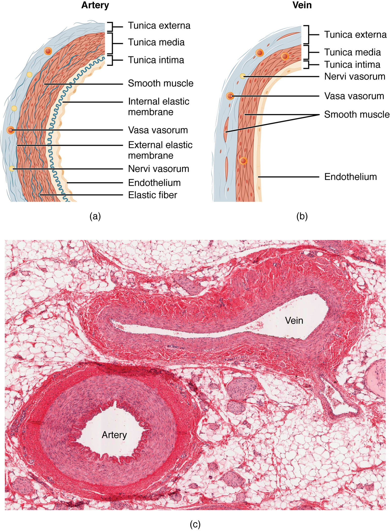
Arteries Arteries are blood vessels with thick, muscular walls designed to withstand the high pressure of blood pumped from the heart. These walls consist of three layers—tunica intima, tunica media, and tunica adventitia—with the tunica media being particularly robust to handle the pulsatile flow.
Veins Veins have thinner walls compared to arteries, as they transport blood back to the heart under lower pressure. Their structure includes valves to prevent backflow, with a less prominent tunica media, accommodating the slower, steadier blood movement.
Micrograph Micrograph offers a 160x magnified view, revealing the relative thickness of arterial and venous walls, providing a clear contrast between the two. This image, provided by the Regents of the University of Michigan Medical School, aids in understanding the histological differences under a light microscope.
The Role of Blood Vessel Structure in Circulation
This stage of exploration delves into how blood vessels are tailored to their functions. The differences in wall thickness directly influence their ability to manage blood pressure and flow.
- Arterial Design: The thick tunica media in arteries contains smooth muscle and elastic fibers, allowing them to expand and recoil with each heartbeat. This elasticity helps maintain blood pressure during diastole, ensuring continuous flow to tissues.
- Venous Adaptation: Veins rely on a thinner tunica media and skeletal muscle action to return blood to the heart against gravity. The presence of valves ensures unidirectional flow, preventing pooling in the lower extremities.
- Microscopic Insight: The micrograph highlights the dense elastic lamellae in arteries versus the looser connective tissue in veins. This contrast is critical for diagnosing vascular conditions through histological analysis.
Anatomical Layers of Blood Vessels
The image provides a detailed sectional view, showcasing the layered construction of arteries and veins. These layers work together to support the vessel’s specific role in the circulatory system.
Arteries and veins share a similar three-layered structure, though their proportions vary significantly. The microscopic view enhances understanding of how these layers adapt to their environments.
- Tunica Intima: This inner layer, lined with endothelium, reduces friction and facilitates blood flow in both arteries and veins. In arteries, it is supported by a thin elastic lamina, while in veins, it is less rigid.
- Tunica Media: The middle layer in arteries is thick with smooth muscle and elastic fibers, enabling vasoconstriction and vasodilation. In veins, this layer is thinner, reflecting the lower pressure they endure.
- Tunica Adventitia: The outer layer provides structural support with collagen and connective tissue, anchoring vessels to surrounding tissues. Veins have a relatively thicker adventitia to compensate for their thinner media.
- Valves in Veins: Unique to veins, these flap-like structures prevent backflow, especially in the lower body. They are absent in arteries due to the high, continuous pressure.
Physiological Functions of Arteries and Veins
The physiological roles of these vessels are shaped by their anatomical differences. Their design ensures efficient blood distribution and return throughout the body.
Arteries actively propel blood, while veins passively return it, creating a balanced circulatory system. The micrograph underscores how these functions are reflected at a cellular level.
- Arterial Blood Pressure: The thick muscular walls of arteries withstand systolic pressure, up to 120 mmHg in healthy adults. Elastic recoil during diastole maintains pressure, supporting peripheral circulation.
- Venous Return: Veins operate at a lower pressure, around 10-15 mmHg, relying on the respiratory pump and muscle contraction. Valves ensure blood moves toward the heart despite gravity.
- Oxygen and Nutrient Delivery: Arteries carry oxygenated blood from the heart, rich in nutrients like glucose and oxygen. Veins return deoxygenated blood, carrying carbon dioxide and waste products.
- Microcirculation Link: The transition from arteries to capillaries and back to veins involves pressure drops, managed by the vessel wall thickness. This gradient supports exchange in capillary beds.
Comparative Histology Under the Microscope
The micrograph provides a unique perspective on vessel wall composition. The 160x magnification reveals details not visible to the naked eye, enhancing anatomical study.
This high-resolution view allows for a deeper understanding of tissue organization. It serves as a valuable tool for exploring vascular health.
- Arterial Wall Thickness: The micrograph shows a prominent tunica media, with multiple elastic lamellae visible. This thickness correlates with the artery’s role in high-pressure environments.
- Venous Wall Thinness: The thinner tunica media in veins is evident, with more collagen in the adventitia. This structure supports flexibility and valve function.
- Cellular Details: Endothelial cells line both vessel types, but arterial cells are more elongated due to pressure. Venous endothelium appears more relaxed, reflecting lower stress.
- Staining Techniques: The micrograph likely uses hematoxylin and eosin staining, highlighting elastic fibers in arteries and connective tissue in veins. This technique aids in differentiating the two.
Clinical and Research Perspectives
Understanding blood vessel anatomy has significant implications for medical practice. The differences between arteries and veins are critical for diagnosing and treating vascular diseases.
Research continues to explore these structures, offering insights into circulatory health. Advanced imaging and histological studies enhance these efforts.
- Atherosclerosis Risk: Thick arterial walls are prone to plaque buildup, narrowing the lumen. Early detection through microscopy can guide preventive measures.
- Varicose Veins: Weak venous walls and valve incompetence lead to vein dilation. Micrographs help assess structural integrity in affected patients.
- Surgical Applications: Knowledge of vessel layers informs procedures like bypass grafting. Arterial thickness requires specific suturing techniques.
- Therapeutic Innovations: Targeting tunica media smooth muscle can treat hypertension. Stem cell research explores regenerating damaged vessel walls.
In conclusion, this image of blood vessel sectional and microscopic views offers a comprehensive look at the structural differences between arteries and veins, essential for their circulatory roles. These insights not only deepen our understanding of vascular anatomy but also support advancements in medical diagnosis and treatment.

