The cerebral cortex is a marvel of the human brain, orchestrating a complex array of sensory, motor, and integrative functions that define our interactions with the world. This diagram illustrates the distinct types of cortical areas—primary, association, and integration regions—each playing a unique role in processing information and coordinating responses. Understanding these regions provides a foundation for appreciating how the brain interprets sensory input and executes motor commands, making this a vital topic for those interested in neuroscience.
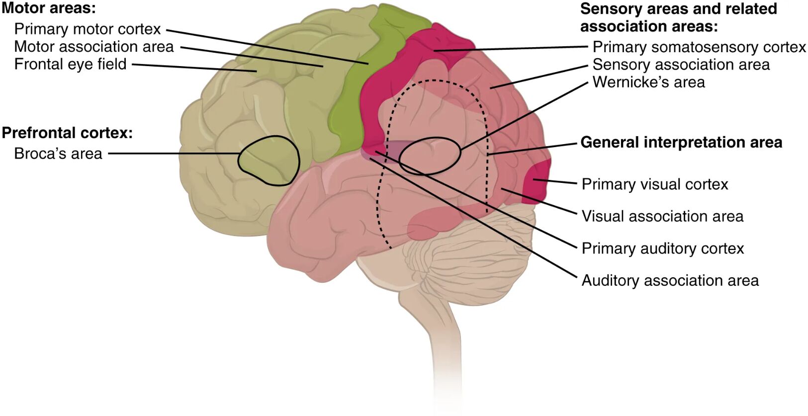
Primary motor cortex The primary motor cortex, located in the frontal lobe, is responsible for initiating voluntary movements by sending signals to the muscles via the corticospinal tract. It contains a topographic map, known as the motor homunculus, where different body parts are represented based on their motor control needs.
Motor association area The motor association area, also in the frontal lobe, plans and coordinates complex movements by integrating information from other brain regions. It works closely with the primary motor cortex to refine motor sequences before execution.
Frontal eye field The frontal eye field, situated in the frontal cortex, controls voluntary eye movements and gaze direction, enabling visual exploration and tracking. It integrates with visual and motor systems to adjust eye position in response to stimuli.
Broca’s area Broca’s area, located in the prefrontal cortex, is critical for speech production and language articulation, coordinating the muscles involved in speaking. Damage here can lead to expressive aphasia, impairing the ability to form coherent sentences.
Prefrontal cortex The prefrontal cortex governs higher cognitive functions such as decision-making, personality expression, and planning, acting as the brain’s executive center. It modulates emotional responses and long-term memory through connections with other cortical areas.
General interpretation area The general interpretation area, found at the junction of multiple cortical regions, integrates multimodal sensory information for complex thought and problem-solving. It enables the brain to synthesize data from vision, hearing, and touch into a coherent understanding.
Primary somatosensory cortex The primary somatosensory cortex, located in the parietal lobe, processes tactile sensations like touch, pressure, and pain from the body’s surface. It features a sensory homunculus, mapping body parts according to their sensory sensitivity.
Sensory association area The sensory association area, adjacent to the primary somatosensory cortex, interprets and integrates tactile data into meaningful perceptions, such as recognizing object shapes. It enhances the brain’s ability to process spatial and temporal aspects of touch.
Wernicke’s area Wernicke’s area, situated in the temporal lobe, is essential for language comprehension and understanding spoken or written words. Damage to this region can result in receptive aphasia, where individuals struggle to understand language.
Primary visual cortex The primary visual cortex, located in the occipital lobe, receives and processes initial visual input from the retina via the optic nerve. It detects basic features like edges and motion, forming the foundation for visual perception.
Visual association area The visual association area, surrounding the primary visual cortex, interprets visual data to recognize objects, faces, and patterns. It builds on raw visual input to create a detailed and contextual understanding of the environment.
Primary auditory cortex The primary auditory cortex, found in the temporal lobe, processes basic sound frequencies and tones received from the cochlea. It serves as the initial stage for auditory perception, distinguishing pitch and volume.
Auditory association area The auditory association area, adjacent to the primary auditory cortex, interprets sounds to recognize speech, music, or environmental noises. It integrates auditory input with memory to facilitate language and sound recognition.
Understanding the Primary Cortical Areas
Primary cortical areas are the first point of contact for sensory or motor signals. This diagram highlights their foundational role in neural processing.
- The primary motor cortex activates specific muscle groups, with larger representations for hands and face due to their dexterity.
- The primary somatosensory cortex maps the body’s sensory input, with heightened sensitivity in areas like the fingertips.
- The primary visual cortex processes raw visual data, sending it to higher areas for interpretation.
- The primary auditory cortex decodes sound waves, enabling basic hearing capabilities.
- These regions rely on direct neural pathways, making them vulnerable to localized damage.
Role of Association Areas in Sensory Integration
Association areas enhance the brain’s ability to interpret sensory data beyond initial processing. Their proximity to primary areas fosters complex analysis.
- The sensory association area refines touch into perceptions like texture or temperature gradients.
- Wernicke’s area decodes language, linking sounds to meaning for comprehension.
- The visual association area recognizes familiar objects by comparing new input with stored memories.
- The auditory association area distinguishes voices or melodies, aiding communication.
- These regions amplify sensory detail, supporting advanced cognitive tasks.
Function of the General Interpretation Area
The general interpretation area serves as a hub for multimodal integration. Its location at cortical junctions enables broad functional synthesis.
- This area combines visual and auditory data to understand spoken instructions.
- It processes spatial relationships across senses, such as locating a sound’s source.
- The region supports abstract thinking, like solving puzzles using multiple inputs.
- It interacts with the prefrontal cortex for decision-making based on integrated data.
- Damage here can disrupt overall perception, affecting daily activities.
Anatomical Organization of the Prefrontal Cortex
The prefrontal cortex orchestrates higher-order functions critical for behavior and cognition. This diagram underscores its diverse roles.
- Broca’s area coordinates speech muscles, essential for fluent verbal communication.
- The prefrontal cortex plans future actions, relying on working memory networks.
- It regulates impulse control, influencing social and emotional responses.
- Connections with the limbic system modulate mood via serotonin and dopamine.
- Thyroid hormones like T3 and T4 enhance its metabolic activity, impacting focus.
Clinical Relevance of Cortical Mapping
Understanding cortical areas aids in diagnosing and treating neurological conditions. This visual guide supports clinical applications.
- Lesions in the primary motor cortex can cause paralysis, assessed via strength tests.
- Damage to Wernicke’s area may lead to language comprehension deficits.
- Visual cortex injuries result in blind spots or hallucinations, detectable via perimetry.
- Auditory cortex damage can cause hearing loss, evaluated with audiometry.
- Imaging like fMRI maps these areas to guide surgical interventions.
Impact of Cortical Damage on Function
Cortical damage disrupts specific functions, with effects varying by region. This diagram provides a basis for predicting outcomes.
- Stroke in the motor association area may impair movement planning, leading to clumsiness.
- Injury to the sensory association area can hinder object recognition via touch.
- Visual association damage might cause inability to interpret faces, known as prosopagnosia.
- Auditory association issues can affect music appreciation or speech understanding.
- Rehabilitation focuses on compensatory mechanisms in undamaged areas.
Advances in Cortical Research
Ongoing studies enhance our knowledge of cortical functions. This diagram inspires exploration into neural plasticity.
- Neuroimaging tracks cortical changes during learning or recovery.
- Transcranial magnetic stimulation modulates activity in specific areas.
- Research on association areas reveals their role in creativity and problem-solving.
- Genetic studies explore how cortical development influences function.
- These advances inform therapies for conditions like aphasia or motor deficits.
In conclusion, this diagram of the cerebral cortex’s types of areas offers a comprehensive view of how the brain processes sensory input, plans movements, and integrates information. From the primary motor and sensory regions to the sophisticated association and interpretation areas, each plays a vital role in human function. This understanding not only deepens appreciation for neural complexity but also supports advancements in diagnosing and treating related disorders, making it an invaluable resource for exploring brain health.

