The central nervous system (CNS) serves as the body’s command center, orchestrating a wide range of functions that can be assessed through a structured neurological exam. This diagram provides a clear visual representation of how different CNS regions correlate with key components of the exam, including mental status, cranial nerves, motor skills, sensory perception, and coordination. By examining these anatomical underpinnings, one can gain a deeper understanding of how the brain and spinal cord interact to maintain normal physiological functions and respond to clinical evaluation.
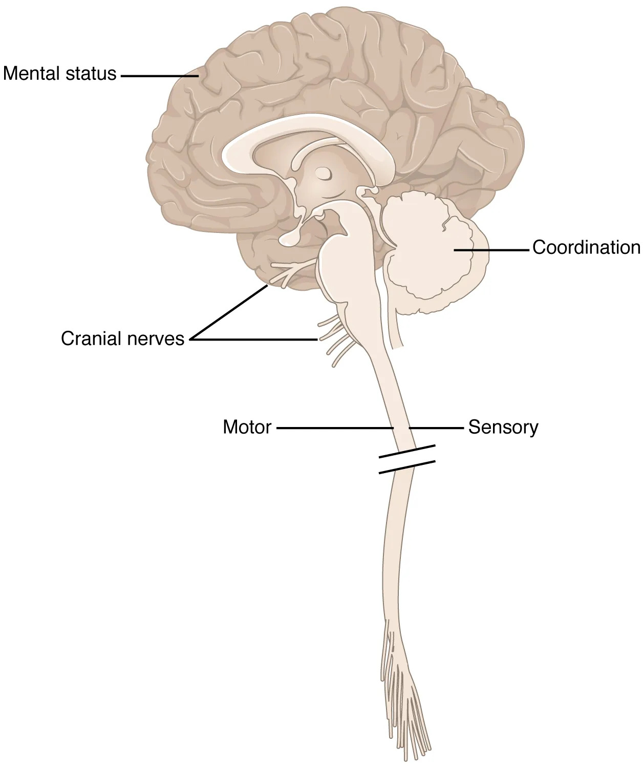
Mental status Mental status reflects the brain’s cognitive and emotional functions, assessed through tests of memory, attention, and mood. This region, primarily the cerebral cortex, integrates higher-order processes essential for consciousness and decision-making.
Coordination Coordination involves the cerebellum, located at the back of the brain, which fine-tunes movement and balance. Its role is critical in tasks like walking or pointing, ensuring smooth and precise motor actions.
Cranial nerves Cranial nerves emerge from the brainstem and control functions such as facial movement, vision, and hearing, with twelve pairs each serving specific sensory or motor roles. Their assessment helps identify localized neurological deficits or damage.
Motor The motor region, spanning the cerebral cortex and spinal cord, governs voluntary muscle movements through upper and lower motor neurons. It enables actions like lifting an arm, with disruptions indicating potential nerve or muscle issues.
Sensory The sensory area, involving the spinal cord and sensory cortex, processes touch, pain, and temperature from the body’s periphery. This region’s evaluation reveals how effectively the nervous system relays external stimuli to the brain.
Overview of the Central Nervous System in Neurological Assessment
The CNS forms the foundation of the neurological exam, with each region contributing uniquely. This diagram highlights the interconnected roles of these areas in daily function.
- The central nervous system comprises the brain and spinal cord, housing billions of neurons that coordinate bodily activities.
- The cerebral cortex, linked to mental status, processes complex thought and emotional regulation.
- The brainstem, home to cranial nerve nuclei, bridges the brain and spinal cord, managing vital reflexes.
- The spinal cord segments handle motor and sensory signals, transmitting them bidirectionally.
- The cerebellum’s coordination role ensures fluid movement, often tested through heel-to-shin tasks.
Role of the Cerebral Cortex in Mental Status Evaluation
The cerebral cortex is pivotal for cognitive functions assessed in the mental status exam. Its layered structure supports diverse mental activities.
- This region includes the frontal lobe, which governs reasoning and problem-solving.
- The parietal lobe processes spatial awareness and sensory integration.
- Memory relies on the temporal lobe, where hippocampal structures store and retrieve information.
- Emotional responses are modulated by the limbic system, including the amygdala.
- Deficits here might manifest as confusion or impaired judgment during exams.
Cranial Nerves and Their Clinical Significance
Cranial nerves offer a direct window into brainstem function and peripheral nerve health. Their assessment is a key component of the neurological exam.
- The olfactory nerve (I) detects smells, often tested with familiar scents.
- The optic nerve (II) carries visual data, evaluated via visual acuity tests.
- The oculomotor (III), trochlear (IV), and abducens (V) nerves control eye movement and pupil response.
- The facial nerve (VII) manages facial expressions, tested by smiling or frowning.
- Damage to these nerves can indicate conditions like Bell’s palsy or brainstem lesions.
Motor and Sensory Pathways in the Spinal Cord
The spinal cord serves as a conduit for motor and sensory information, critical for movement and perception. This diagram emphasizes their distinct yet overlapping roles.
- Motor pathways originate in the motor cortex, descending via the corticospinal tract to activate muscles.
- Lower motor neurons in the spinal cord connect to muscles, enabling reflexes like the knee-jerk response.
- Sensory pathways ascend through the dorsal columns and spinothalamic tracts to the brain.
- Touch, pain, and temperature are relayed via different fiber types, tested with pinpricks or vibration.
- Lesions here can lead to weakness or numbness, guiding diagnostic imaging.
Cerebellar Function and Coordination Testing
The cerebellum fine-tunes movements, making coordination a vital exam component. Its anatomical position supports precise motor control.
- The cerebellar hemispheres coordinate voluntary movements like finger-to-nose tests.
- The vermis regulates axial movements, such as maintaining posture during walking.
- Purkinje cells within the cerebellum integrate sensory input for balance.
- Ataxia, or lack of coordination, signals cerebellar dysfunction, often assessed via gait.
- Alcohol or stroke can impair this region, affecting fine motor skills.
Clinical Applications of the Neurological Exam
This diagram aids in applying the neurological exam to diagnose and monitor conditions. It links anatomy to practical assessment techniques.
- Mental status changes might suggest dementia or delirium, requiring cognitive testing.
- Cranial nerve palsies can point to tumors or multiple sclerosis affecting the brainstem.
- Motor weakness may indicate stroke or spinal cord injury, assessed with strength grading.
- Sensory loss could stem from peripheral neuropathy or spinal compression.
- Coordination issues guide MRI scans to evaluate cerebellar or vestibular pathology.
Neurotransmitters and Hormonal Influences
Chemical signaling within the CNS enhances the exam’s interpretive value. These substances modulate neural activity.
- Acetylcholine supports memory and motor function, affected in Alzheimer’s disease.
- Dopamine regulates movement, with deficits linked to Parkinson’s disease.
- Serotonin influences mood, assessed in mental status evaluations.
- Thyroid hormones like T3 and T4 impact overall neural metabolism and responsiveness.
- Imbalances can alter exam findings, necessitating hormonal screening.
Advances in Neurological Imaging and Assessment
Modern technology complements this anatomical framework, enhancing exam precision. The diagram inspires further exploration.
- MRI and CT scans visualize structural changes in the brain and spinal cord.
- Electroencephalography (EEG) monitors electrical activity for seizure detection.
- Functional MRI (fMRI) maps brain regions during cognitive tasks.
- Nerve conduction studies assess peripheral nerve integrity.
- These tools refine diagnoses based on exam findings, improving patient outcomes.
In conclusion, this diagram of the anatomical underpinnings of the neurological exam offers a comprehensive view of how the central nervous system’s regions contribute to clinical assessments. From mental status to coordination, each area plays a distinct role in maintaining bodily function, with the exam serving as a critical tool to detect abnormalities. This visual resource not only enhances understanding of neural anatomy but also supports ongoing efforts to refine diagnostic and therapeutic approaches in neurology.

