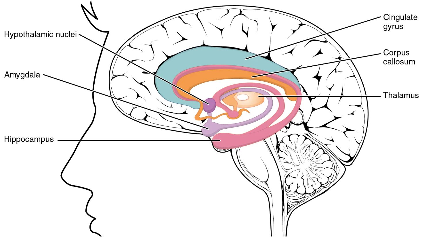
The limbic lobe structure diagram offers a detailed view of the brain regions that play a pivotal role in emotion, memory, and autonomic regulation, forming a critical part of the brain’s inner architecture. This chart highlights key components such as the amygdala, hippocampus, and cingulate gyrus, which encircle the cerebrum and connect to the hypothalamus, influencing both conscious and unconscious behaviors. Exploring this structure provides valuable insights into the neural basis of human experience and physiological control.
Labeled Components in the Diagram
Hypothalamic nuclei The hypothalamic nuclei are clusters of cells within the hypothalamus that regulate vital functions like hunger, thirst, and hormone release. They connect with the limbic lobe to coordinate emotional responses with bodily states.
Amygdala The amygdala is a small almond-shaped structure involved in processing emotions, particularly fear and pleasure. It interacts with the hypothalamus to trigger autonomic responses to emotional stimuli.
Hippocampus The hippocampus plays a central role in forming and retrieving memories, located within the medial temporal lobe. It collaborates with other limbic structures to integrate spatial and episodic memory.
Cingulate gyrus The cingulate gyrus is a curved fold of the cerebral cortex above the corpus callosum, linked to emotion regulation and pain perception. It facilitates communication between the limbic system and the prefrontal cortex.
Corpus callosum The corpus callosum is a thick band of nerve fibers that connects the left and right hemispheres, enabling interhemispheric communication. It supports the integration of limbic functions across both brain halves.
Thalamus The thalamus acts as a relay station, transmitting sensory and motor signals to the cerebral cortex and linking with limbic structures. It modulates the flow of information to and from the limbic lobe.
Anatomy of the Limbic Lobe
The limbic lobe forms a ring-like structure around the brainstem and corpus callosum, deeply embedded in the brain’s emotional and memory systems. This region bridges the cerebral cortex with subcortical areas for integrated function.
- The hippocampus and amygdala reside in the medial temporal lobe, critical for memory and emotion.
- The cingulate gyrus extends over the corpus callosum, influencing emotional processing.
- The hypothalamic nuclei connect the limbic lobe to endocrine regulation.
- The thalamus serves as a gateway, relaying sensory input to limbic regions.
- The corpus callosum ensures synchronized activity between hemispheres.
- These structures collectively form the limbic system, vital for survival instincts.
Physiological Roles of Limbic Structures
Each component of the limbic lobe contributes to specific physiological processes, maintaining a balance between emotion and bodily function. This coordination is essential for adaptive behavior.
- The amygdala releases stress hormones like cortisol during fear responses.
- The hippocampus generates new neurons in the dentate gyrus for memory consolidation.
- The cingulate gyrus modulates heart rate and blood pressure via autonomic links.
- The hypothalamic nuclei secrete releasing hormones such as CRH and TRH.
- The thalamus filters sensory data, prioritizing signals for limbic processing.
- The corpus callosum synchronizes emotional experiences across hemispheres.
Emotional Regulation and the Limbic Lobe
The limbic lobe is a cornerstone of emotional regulation, influencing how individuals respond to their environment. Its connectivity enhances adaptive emotional responses.
- The amygdala triggers the fight-or-flight response through the sympathetic nervous system.
- The hippocampus links emotions to specific memories, shaping emotional context.
- The cingulate gyrus mediates conflict resolution and empathy.
- The hypothalamic nuclei adjust mood through hormone release like oxytocin.
- The thalamus relays emotional cues from sensory inputs.
- The corpus callosum balances emotional expression between brain hemispheres.
Memory Function and the Hippocampus
The hippocampus stands out for its role in memory formation and spatial navigation. Its interactions with other limbic structures enhance cognitive abilities.
- It processes short-term memories into long-term storage.
- The hippocampus collaborates with the amygdala to encode emotional memories.
- Spatial memory relies on its connection to the entorhinal cortex.
- Neurogenesis in the hippocampus supports learning and adaptability.
- Damage can lead to anterograde amnesia, impairing new memory formation.
- Its location near the thalamus aids in sensory memory integration.
Clinical Relevance of Limbic Structures
Understanding the limbic lobe aids in diagnosing and treating neurological and psychiatric conditions. These insights guide therapeutic approaches.
- Amygdala hyperactivity is linked to anxiety disorders and PTSD.
- Hippocampus atrophy occurs in Alzheimer’s disease, affecting memory.
- Cingulate gyrus dysfunction is associated with depression and OCD.
- Hypothalamic nuclei imbalances can cause hormonal disorders like diabetes insipidus.
- Thalamus lesions may result in sensory processing deficits.
- Corpus callosum abnormalities are seen in conditions like agenesis of the corpus callosum.
Advances in Limbic Research
Ongoing research into the limbic lobe offers new perspectives on brain function and potential treatments. These developments hold promise for future healthcare.
- MRI techniques map hippocampus volume changes in dementia.
- Deep brain stimulation targets the cingulate gyrus for OCD treatment.
- Stem cell therapy aims to regenerate hippocampus neurons.
- Pharmacological agents modulate amygdala activity in anxiety.
- Functional connectivity studies explore thalamus-limbic links.
- Virtual reality assesses spatial memory via the hippocampus.
The limbic lobe structure underscores the brain’s remarkable capacity to integrate emotion, memory, and physiological regulation. By delving into these regions, professionals can enhance diagnostic precision and develop innovative therapies, ultimately improving understanding and care for those affected by related conditions.

