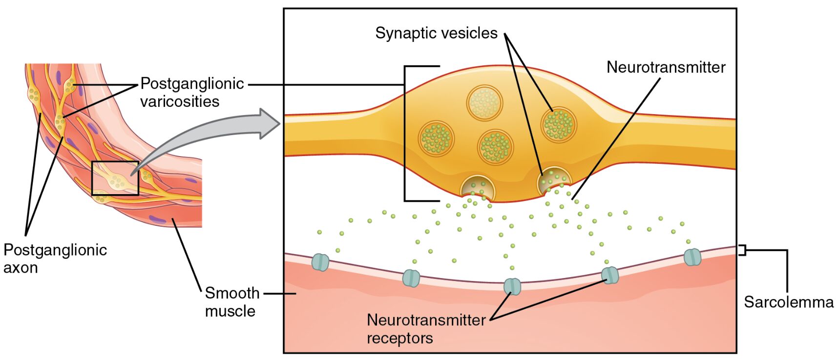
The autonomic nervous system regulates involuntary bodily functions through unique neural connections, differing significantly from the typical neuromuscular junction. This diagram highlights the role of autonomic varicosities, where neurotransmitters are released from swellings along postganglionic fibers to form an extensive network, influencing target effectors like smooth muscle in a diffuse manner.
Labels in the Diagram
Postganglionic Varicosities The Postganglionic Varicosities are swellings along the postganglionic axon where neurotransmitters are released. These structures allow for widespread influence on target tissues, unlike the focused synapse of skeletal muscle.
Postganglionic Axon The Postganglionic Axon extends from the ganglion to the target effector, featuring varicosities along its length. It transmits signals from the autonomic nervous system to smooth muscle or glands.
Smooth Muscle The Smooth Muscle is the target tissue innervated by postganglionic fibers, found in organs like the stomach and blood vessels. It contracts or relaxes in response to neurotransmitter release from varicosities.
Sarcolemma The Sarcolemma is the cell membrane of smooth muscle cells, containing neurotransmitter receptors. It receives and responds to the chemical signals released by varicosities.
Synaptic Vesicles The Synaptic Vesicles within the axon terminal store neurotransmitters, ready for release. They migrate to the varicosity membrane to discharge their contents upon stimulation.
Neurotransmitter The Neurotransmitter is the chemical messenger released from synaptic vesicles into the synaptic cleft. It binds to receptors on the sarcolemma to elicit a response in the smooth muscle.
Neurotransmitter Receptors The Neurotransmitter Receptors are proteins on the sarcolemma that detect and bind neurotransmitters. This binding triggers intracellular changes, leading to muscle contraction or relaxation.
Anatomy of Autonomic Varicosities
The autonomic nervous system employs a distinctive mechanism for communication, relying on postganglionic varicosities rather than traditional synaptic end bulbs. These varicosities, found along the postganglionic axon, release neurotransmitter from synaptic vesicles into a diffuse area surrounding the smooth muscle.
- Varicosity Structure: Swellings contain numerous synaptic vesicles filled with neurotransmitter.
- Axon Pathway: The postganglionic axon forms an extensive network, enhancing coverage of target tissues.
- Muscle Response: The sarcolemma with neurotransmitter receptors mediates the physiological effect.
This diffuse release allows for broader and more flexible control compared to the precise point-to-point synapses of the somatic nervous system.
Physiological Role of Varicosities in Smooth Muscle Control
The postganglionic varicosities enable precise yet widespread regulation of smooth muscle function. Release of neurotransmitter from these sites activates neurotransmitter receptors on the sarcolemma, initiating responses like vasoconstriction or gastrointestinal motility.
- Vasomotor Control: Regulates blood vessel diameter to adjust blood pressure.
- Digestive Motility: Enhances peristalsis in the intestines for digestion.
- Pupil Adjustment: Influences iris muscle to control pupil size in the eye.
This mechanism supports the autonomic system’s role in maintaining homeostasis across various organs.
Comparison with Neuromuscular Junctions
Unlike the neuromuscular junction, where a single synapse delivers a concentrated signal, autonomic varicosities provide a diffuse release pattern. The postganglionic axon with its varicosities releases neurotransmitter over a larger area, engaging multiple neurotransmitter receptors on the sarcolemma.
- Synaptic Efficiency: Neuromuscular junctions offer rapid, targeted responses for skeletal muscle.
- Diffuse Influence: Varicosities allow graded responses suited for smooth muscle regulation.
- Neurotransmitter Dynamics: Continuous release from synaptic vesicles supports sustained effects.
This difference reflects the autonomic system’s need for nuanced control over involuntary functions.
Clinical Relevance of Autonomic Varicosities
Understanding autonomic varicosities is crucial for addressing disorders involving smooth muscle dysfunction. Abnormal neurotransmitter release or receptor sensitivity can lead to conditions like hypertension or irritable bowel syndrome.
- Hypertension: Overactive varicosity release may cause excessive vasoconstriction.
- Gastrointestinal Disorders: Altered neurotransmitter receptors affect motility.
- Therapeutic Targets: Drugs modulating neurotransmitter action can treat these conditions.
Accurate diagnosis relies on recognizing the unique anatomy of these connections.
Evolutionary Perspective and Research Insights
The autonomic varicosities likely evolved to provide flexible control over smooth muscle in early organisms, supporting survival functions like digestion and circulation. Modern research continues to explore their role in health and disease.
- Evolutionary Advantage: Diffuse innervation enhanced adaptability in primitive systems.
- Neuroimaging Studies: Reveal synaptic vesicles distribution in varicosities.
- Therapeutic Potential: Insights guide development of autonomic-targeting medications.
This knowledge deepens our understanding of autonomic regulation and its clinical applications.
In conclusion, autonomic varicosities represent a fascinating adaptation in the autonomic nervous system, enabling effective communication with smooth muscle through a network of postganglionic axons. This diagram offers a clear view of the process, from synaptic vesicles to neurotransmitter receptors, providing a foundation for both anatomical study and medical practice.

