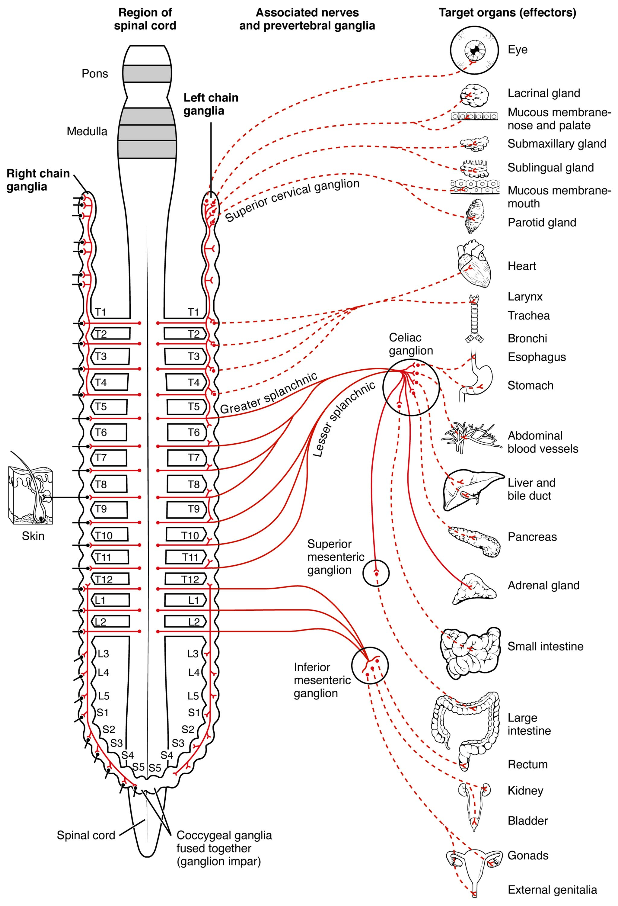
The sympathetic division of the autonomic nervous system plays a vital role in the body’s “fight or flight” response, coordinating rapid physiological changes to handle stress or danger. This diagram provides a comprehensive overview of how preganglionic and postganglionic neurons connect the spinal cord to various target organs, illustrating the intricate network that maintains homeostasis under challenging conditions.
Labels in the Diagram
Pons The Pons is a region of the brainstem that contributes to the regulation of autonomic functions, including parts of the sympathetic nervous system. It serves as a relay station, connecting higher brain centers to the spinal cord for coordinated responses.
Medulla The Medulla, also part of the brainstem, houses critical autonomic control centers that influence heart rate and breathing. It integrates signals from the sympathetic division to maintain vital bodily functions.
Right Chain Ganglia The Right Chain Ganglia are part of the paravertebral ganglia located along the right side of the spinal column. These ganglia receive preganglionic fibers and relay signals to postganglionic neurons targeting various effectors.
Left Chain Ganglia The Left Chain Ganglia, similarly positioned along the left side of the vertebral column, form a symmetrical network with the right chain. They facilitate the distribution of sympathetic signals to the body’s lateral regions.
Superior Cervical Ganglion The Superior Cervical Ganglion is a key paravertebral ganglion located near the upper cervical spine. It innervates structures in the head and neck, such as the eyes and salivary glands, via postganglionic fibers.
Greater Splanchnic Nerve The Greater Splanchnic Nerve carries preganglionic fibers from the thoracic spinal cord to the celiac ganglion. It plays a major role in innervating abdominal organs like the stomach and liver.
Lesser Splanchnic Nerve The Lesser Splanchnic Nerve extends from the lower thoracic levels to the aorticorenal ganglion. It supports sympathetic control over organs such as the kidneys and adrenal glands.
Celiac Ganglion The Celiac Ganglion is a prevertebral ganglion located in the abdominal cavity that receives input from splanchnic nerves. It distributes postganglionic fibers to digestive organs, influencing their activity during stress.
Superior Mesenteric Ganglion The Superior Mesenteric Ganglion, another prevertebral ganglion, regulates blood flow and motility in the small intestine. It receives preganglionic fibers via the splanchnic nerves for sympathetic control.
Inferior Mesenteric Ganglion The Inferior Mesenteric Ganglion controls the large intestine and other pelvic structures. It integrates sympathetic input to modulate gastrointestinal and genitourinary functions.
Coccygeal Ganglia (Ganglion Impar) The Coccygeal Ganglia, or ganglion impar, is a fused ganglion at the base of the spinal cord. It provides sympathetic innervation to the pelvic region, including the external genitalia.
Spinal Cord The Spinal Cord serves as the origin of preganglionic neurons in the lateral horn, particularly from thoracic (T1-T12) and upper lumbar (L1-L2) segments. These neurons project to ganglia to initiate the sympathetic response.
Skin The Skin receives sympathetic innervation that regulates sweat gland activity and blood vessel constriction. This helps in thermoregulation and response to environmental stress.
Eye The Eye is innervated by the superior cervical ganglion, controlling pupil dilation via the radial muscle. This response enhances visual acuity during heightened alertness.
Lacrimal Gland The Lacrimal Gland produces tears and is influenced by sympathetic stimulation to adjust tear production. This helps maintain eye moisture during stress responses.
Mucous Membrane – Nose and Palate The Mucous Membrane – Nose and Palate receives sympathetic input to reduce secretions, aiding in airway clearance during the fight-or-flight response.
Submaxillary Gland The Submaxillary Gland, a salivary gland, is modulated by sympathetic nerves to decrease saliva production. This prepares the body by redirecting resources during stress.
Sublingual Gland The Sublingual Gland similarly reduces saliva output under sympathetic control. This conserves energy for more immediate survival needs.
Mucous Membrane – Mouth The Mucous Membrane – Mouth experiences reduced secretions to minimize oral moisture. This supports the body’s focus on critical functions during stress.
Parotid Gland The Parotid Gland, another salivary gland, decreases secretion under sympathetic influence. This aligns with the body’s shift away from non-essential activities.
Heart The Heart receives sympathetic input to increase heart rate and contractility. This ensures adequate blood flow to muscles during emergencies.
Larynx The Larynx is modulated to adjust vocal cord tension, aiding in breathing or vocalization during stress. Sympathetic activation prepares it for rapid air exchange.
Trachea The Trachea dilates under sympathetic control to improve airflow to the lungs. This enhances oxygen delivery during physical exertion.
Bronchi The Bronchi also dilate to maximize air intake, supporting increased respiratory demand. This is a key adaptation in the sympathetic response.
Esophagus The Esophagus reduces motility under sympathetic influence, pausing digestion. This prioritizes energy for immediate survival needs.
Stomach The Stomach decreases acid secretion and motility during sympathetic activation. This conserves energy for other bodily demands.
Abdominal Blood Vessels The Abdominal Blood Vessels constrict to redirect blood to skeletal muscles. This enhances physical performance during stress.
Liver and Bile Duct The Liver and Bile Duct are stimulated to release glucose into the bloodstream. This provides quick energy for the fight-or-flight response.
Pancreas The Pancreas reduces insulin release under sympathetic control. This helps maintain elevated blood glucose levels for energy.
Adrenal Gland The Adrenal Gland releases adrenaline (epinephrine) in response to sympathetic stimulation. This hormone amplifies the overall stress response.
Small Intestine The Small Intestine slows digestion to conserve energy during sympathetic activation. This redirects resources to critical systems.
Large Intestine The Large Intestine also reduces motility, pausing non-essential processes. This supports the body’s focus on immediate survival.
Rectum The Rectum experiences decreased activity to halt waste elimination. This prevents unnecessary energy expenditure during stress.
Kidney The Kidney reduces urine production under sympathetic influence. This helps maintain blood volume for circulation.
Bladder The Bladder contracts its sphincter to retain urine, aiding focus during emergencies. This is part of the body’s stress adaptation.
Gonads The Gonads reduce activity to conserve energy during sympathetic response. This prioritizes immediate survival over reproduction.
External Genitalia The External Genitalia experience reduced blood flow to focus resources elsewhere. This supports the body’s fight-or-flight priorities.
Anatomy of the Sympathetic Nervous System
The sympathetic division originates in the thoracic and lumbar regions of the spinal cord, where preganglionic neurons exit via the lateral horn. These neurons synapse in chain ganglia or prevertebral ganglia, such as the celiac ganglion, before postganglionic fibers reach target organs.
- Preganglionic Neurons: Short fibers that release acetylcholine to activate ganglia.
- Postganglionic Neurons: Longer fibers that release norepinephrine to stimulate effectors.
- Chain Ganglia: Located along the vertebral column, they form a bilateral network.
This system ensures rapid responses by bypassing lengthy neural pathways, making it efficient for survival.
Physiological Functions of Sympathetic Activation
The sympathetic division prepares the body for stress by accelerating heart rate and dilating airways. This response involves the heart, trachea, and bronchi, enhancing oxygen and nutrient delivery.
- Cardiovascular Effects: Increases blood pressure by constricting abdominal blood vessels.
- Respiratory Effects: Dilates bronchi for improved oxygenation.
- Metabolic Effects: Stimulates the liver to release glucose and the adrenal gland to secrete adrenaline.
These changes collectively support physical exertion and mental alertness during emergencies.
Clinical Relevance and Sympathetic Disorders
Disruptions in the sympathetic division can lead to conditions like hyperhidrosis, excessive sweating due to overactive skin innervation. Understanding these pathways aids in diagnosing and treating autonomic imbalances.
- Hyperhidrosis: Overactivity of sweat glands linked to chain ganglia dysfunction.
- Orthostatic Hypotension: Impaired abdominal blood vessel control causing dizziness.
- Therapeutic Approaches: Medications like beta-blockers modulate heart and lungs responses.
Early intervention can mitigate symptoms, emphasizing the importance of this system’s balance.
Evolutionary Role and Modern Applications
The sympathetic division evolved to enhance survival in predatory environments, coordinating rapid responses via the spinal cord and ganglia. Today, it informs medical fields like anesthesiology and neurology.
- Evolutionary Benefit: Quick mobilization of energy from the liver and pancreas.
- Medical Use: Sympathetic blocks target celiac ganglion for pain management.
- Research Focus: Studying adrenal gland responses improves stress-related treatments.
This knowledge continues to shape clinical practices and patient care.
In conclusion, the sympathetic division of the autonomic nervous system exemplifies the body’s remarkable ability to adapt to stress. This diagram offers a clear roadmap of its connections, from the spinal cord to diverse target organs, providing a foundation for understanding its critical role in health and disease.

