The spinal cord serves as a vital conduit for sensory information, channeling signals from the periphery to the brain through key pathways. This anterior view highlights the dorsal column system and spinothalamic tract, offering a clear perspective on how touch, pressure, vibration, pain, and temperature are transmitted to the central nervous system.
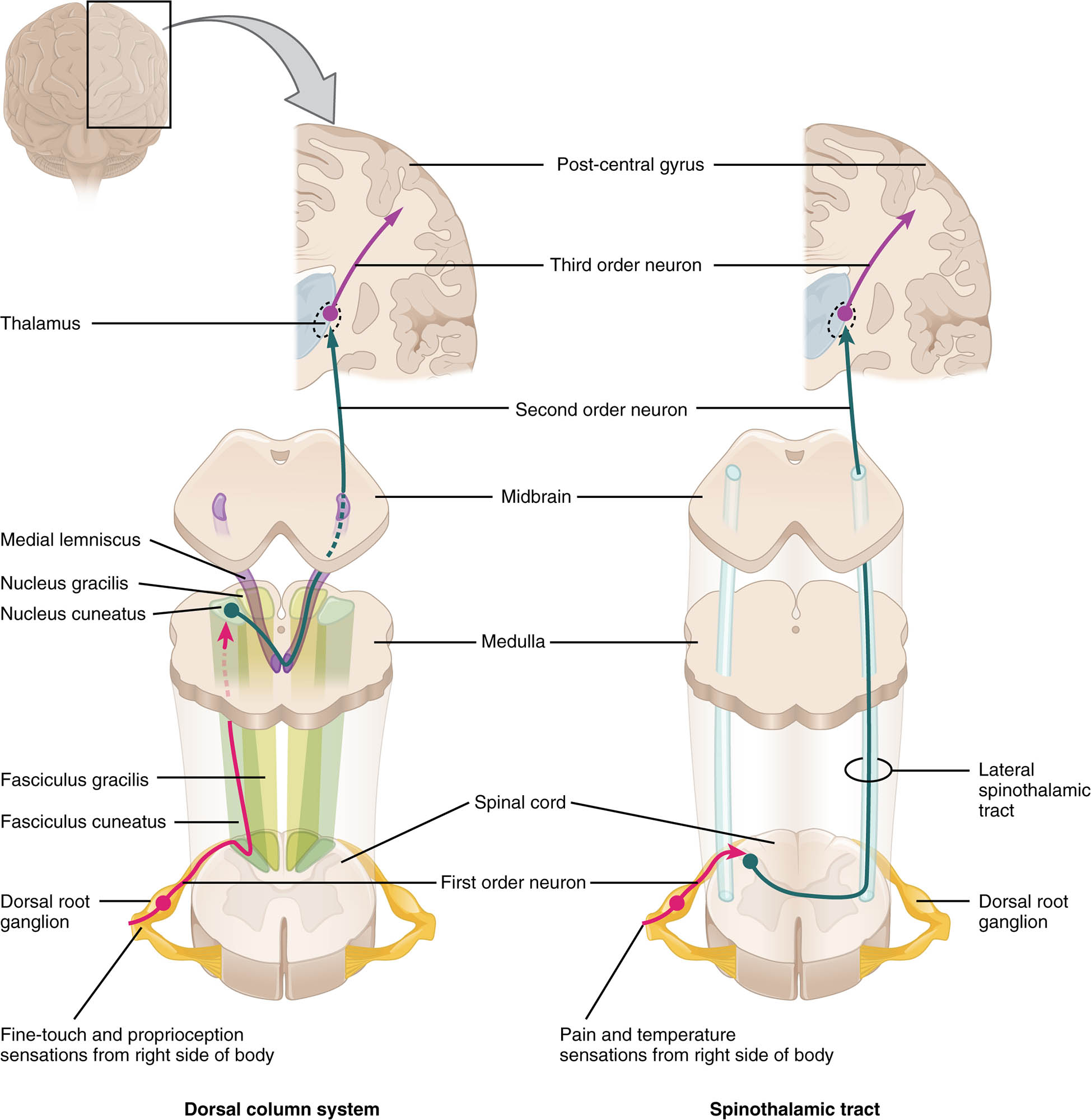
Dorsal root ganglion The dorsal root ganglion contains cell bodies of sensory neurons, serving as the entry point for afferent signals from the periphery. It relays these impulses to the spinal cord for further processing and transmission.
Dorsal column The dorsal column is a major ascending pathway that carries fine touch, vibration, and proprioception signals to the brain. It consists of the fasciculus gracilis and fasciculus cuneatus, which ascend ipsilaterally before synapsing in the medulla.
Fasciculus gracilis The fasciculus gracilis transmits sensory information from the lower body and lower limbs through the dorsal column. It carries signals for touch and proprioception, synapsing in the nucleus gracilis of the medulla oblongata.
Fasciculus cuneatus The fasciculus cuneatus conveys sensory data from the upper body and upper limbs via the dorsal column. It synapses in the nucleus cuneatus, contributing to precise sensory mapping in the brain.
Medulla oblongata The medulla oblongata is where second-order neurons of the dorsal column system cross over, forming the medial lemniscus. It processes and relays sensory information to higher brain centers like the thalamus.
Medial lemniscus The medial lemniscus is the pathway where second-order neurons ascend after decussation in the medulla, carrying fine touch and proprioception to the thalamus. It maintains a somatotopic organization for accurate sensory localization.
Thalamus The thalamus acts as a relay station, receiving sensory inputs from the medial lemniscus and spinothalamic tract. It processes and forwards these signals to the somatosensory cortex for conscious perception.
Spinothalamic tract The spinothalamic tract is an ascending pathway that transmits pain, temperature, and crude touch sensations to the brain. Its fibers decussate within the spinal cord before ascending to the thalamus.
Lateral spinothalamic tract The lateral spinothalamic tract specifically carries pain and temperature sensations, crossing to the opposite side of the spinal cord. It synapses in the thalamus, contributing to the perception of these modalities.
Anterior spinothalamic tract The anterior spinothalamic tract conveys crude touch and pressure sensations, also decussating within the spinal cord. It works alongside the lateral tract to provide a complete sensory profile.
Spinal cord The spinal cord is the central structure where sensory pathways like the dorsal column and spinothalamic tract originate and ascend. It integrates and transmits signals from peripheral nerves to the brain.
Anatomy of Ascending Sensory Pathways
The spinal cord houses the dorsal column system and spinothalamic tract, forming the primary routes for sensory information. This anterior view illustrates their organization and connectivity within the central nervous system.
- The dorsal root ganglion marks the entry of sensory neurons into the spinal cord.
- The dorsal column, divided into fasciculus gracilis and fasciculus cuneatus, handles discriminative touch.
- The spinothalamic tract, including lateral spinothalamic tract and anterior spinothalamic tract, processes pain and touch.
- The medulla oblongata facilitates decussation, ensuring contralateral sensory representation.
- The medial lemniscus carries refined signals to the thalamus for further processing.
- The spinal cord serves as the foundation, with gray and white matter organizing these pathways.
Physiology of Sensory Transmission
Sensory signals travel through the dorsal column system and spinothalamic tract, each with distinct roles in perception. This diagram showcases the physiological flow from periphery to brain.
- The dorsal root ganglion generates action potentials from peripheral stimuli like touch.
- The fasciculus gracilis and fasciculus cuneatus maintain ipsilateral transmission until the medulla oblongata.
- The medial lemniscus relays precise localization to the thalamus after crossing over.
- The lateral spinothalamic tract carries pain and temperature, decussating within the spinal cord.
- The anterior spinothalamic tract transmits crude touch, complementing the lateral tract.
- The thalamus integrates these inputs, sending them to the cortex for conscious awareness.
Role of the Dorsal Column System
The dorsal column system specializes in fine sensory discrimination, relying on its structured pathways. Its anatomy supports detailed perception of the body’s position and texture.
- The fasciculus gracilis handles lower body sensations, preserving spatial accuracy.
- The fasciculus cuneatus processes upper body signals, enhancing hand and arm sensitivity.
- The dorsal column maintains ipsilateral signals, crossing only at the medulla oblongata.
- Second-order neurons form the medial lemniscus, ensuring precise thalamic delivery.
- Damage to this system can lead to loss of vibration sense, though this image shows normal anatomy.
- Proprioception relies heavily on this pathway for coordinated movement.
Role of the Spinothalamic Tract
The spinothalamic tract is essential for detecting pain and temperature, with rapid transmission to the brain. Its design ensures quick responses to potentially harmful stimuli.
- The lateral spinothalamic tract prioritizes pain and temperature, crossing within a few spinal segments.
- The anterior spinothalamic tract adds crude touch, enhancing sensory completeness.
- Decussation in the spinal cord allows contralateral brain representation of these sensations.
- The tract ascends through the anterolateral quadrant, synapsing in the thalamus.
- This pathway’s speed is critical for reflex actions like withdrawing from heat.
- Lesions here can cause dissociated sensory loss, but this diagram depicts healthy structure.
Clinical Relevance of Sensory Pathways
Understanding the dorsal column system and spinothalamic tract aids in diagnosing sensory deficits. This image provides a reference for assessing normal anatomy and function.
- Damage to the dorsal root ganglion can result in sensory neuropathy, affecting all modalities.
- Loss of medial lemniscus function leads to impaired fine touch and proprioception.
- The lateral spinothalamic tract injury causes contralateral pain and temperature loss.
- The anterior spinothalamic tract deficits may reduce crude touch perception.
- Spinal cord injuries disrupting these pathways can lead to sensory ataxia.
- Neuroimaging and sensory testing evaluate pathway integrity for diagnosis.
- Rehabilitation strategies target compensatory mechanisms in pathway damage.
In conclusion, the anterior view of the spinal cord’s ascending sensory pathways reveals a sophisticated network for transmitting touch, pain, and proprioception. The dorsal column system and spinothalamic tract, with their distinct roles and connections, underscore the complexity of sensory processing, offering valuable insights into neurological health.

