Photopigments within the retina are the key to perceiving color, each tuned to specific wavelengths of light that shape our visual experience. This image compares the peak sensitivity and absorbance spectra of these photopigments, offering a detailed look at how they contribute to color vision and visual acuity.
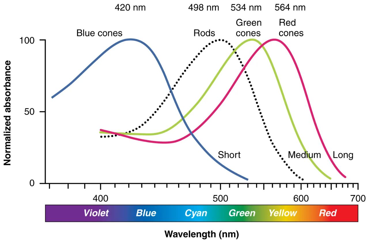
S-cone photopigment The S-cone photopigment is most sensitive to short wavelengths, peaking around 420-440 nm, which corresponds to blue light. It plays a crucial role in detecting blue hues and contributes to color contrast in the visual field.
M-cone photopigment The M-cone photopigment exhibits peak sensitivity to medium wavelengths, approximately 530-540 nm, aligning with green light. This photopigment helps distinguish green shades and supports detailed color perception.
L-cone photopigment The L-cone photopigment is tuned to long wavelengths, with a peak sensitivity around 560-580 nm, corresponding to red light. It is essential for detecting red tones and enhancing color differentiation.
Rod photopigment (rhodopsin) The rod photopigment (rhodopsin) is highly sensitive to a broad range of wavelengths, peaking near 500 nm, and functions best in low-light conditions. Unlike cones, it does not contribute to color vision but is vital for night vision and peripheral sight.
Anatomy of Photopigments in the Retina
Photopigments reside within the photoreceptor cells of the retina, each designed to capture specific light wavelengths. This image illustrates how these pigments’ absorbance spectra define their roles in vision.
- The S-cone photopigment is located in the short-wavelength-sensitive cones, concentrated outside the fovea.
- The M-cone photopigment and L-cone photopigment are found in medium and long-wavelength cones, densely packed in the fovea.
- The rod photopigment (rhodopsin) is housed in rod cells, which outnumber cones and dominate the retina’s periphery.
- These pigments are embedded in the outer segments’ membrane discs or folds, optimizing light absorption.
- The retina’s layered structure positions these photoreceptors to interact with incoming light.
- Supporting cells, like the retinal pigment epithelium, maintain pigment health and regeneration.
Physiology of Color Sensitivity
The absorbance spectra of photopigments determine how the eye perceives color through wavelength-specific sensitivity. This comparison highlights the physiological basis of trichromatic vision.
- The S-cone photopigment detects blue light, contributing to the blue-yellow color axis.
- The M-cone photopigment senses green light, playing a role in the red-green color axis.
- The L-cone photopigment captures red light, complementing the green-sensitive cones.
- The rod photopigment (rhodopsin) responds to a wide spectrum, maximizing sensitivity in dim light.
- When light hits these pigments, it triggers photoisomerization of retinal, initiating a signal.
- The brain integrates signals from all three cone types to perceive the full spectrum of colors.
Role of S-Cone Photopigment in Blue Vision
The S-cone photopigment specializes in short-wavelength detection, crucial for blue light perception. Its unique sensitivity enhances visual contrast in certain lighting conditions.
- The peak at 420-440 nm allows the S-cone photopigment to excel in blue-rich environments.
- This pigment works with opponent processes in the retina to distinguish blue from yellow.
- Its lower density reduces its contribution to overall acuity but aids in color balance.
- Damage to S-cones can lead to tritanopia, a rare blue-yellow color blindness.
- The pigment’s absorbance overlaps minimally with other cones, ensuring distinct blue detection.
- Its role is more pronounced in peripheral vision, complementing central focus.
Role of M- and L-Cone Photopigments in Red-Green Vision
The M-cone photopigment and L-cone photopigment together form the basis of red-green color vision. Their overlapping spectra enable fine color discrimination.
- The M-cone photopigment peaks at 530-540 nm, detecting green and aiding in contrast with red.
- The L-cone photopigment peaks at 560-580 nm, capturing red and enhancing warm tone perception.
- Their close spectral overlap allows the brain to compare signals for color differentiation.
- Anomalies in these pigments cause common color vision deficiencies like deuteranomaly.
- The high concentration in the fovea supports detailed vision and color accuracy.
- These pigments adapt to varying light intensities, maintaining color constancy.
Role of Rod Photopigment in Low-Light Conditions
The rod photopigment (rhodopsin) is designed for broad sensitivity, thriving in low-light environments. Its role complements the cones’ color vision in darker settings.
- The peak at 500 nm makes rod photopigment (rhodopsin) highly effective in dim light.
- This pigment enables scotopic vision, detecting shapes and motion without color.
- Rhodopsin’s high sensitivity allows rod cells to respond to single photons.
- The broad spectrum coverage supports peripheral vision, enhancing night navigation.
- Regeneration of rhodopsin after light exposure takes time, aiding dark adaptation.
- Its function diminishes in bright light, handing over to cone-based vision.
Clinical Relevance of Photopigment Sensitivity
Understanding photopigment absorbance spectra assists in diagnosing and managing vision disorders. This image provides a baseline for exploring related conditions.
- Deficiencies in the S-cone photopigment can lead to tritan defects, affecting blue perception.
- Anomalies in the M-cone photopigment or L-cone photopigment cause red-green color blindness.
- Reduced rod photopigment (rhodopsin) function is linked to night blindness in retinitis pigmentosa.
- Color vision tests, like the Ishihara plates, assess photopigment performance.
- Genetic mutations altering pigment spectra can result in congenital color vision issues.
- Therapies, including gene editing, aim to correct photopigment deficiencies.
- Regular eye exams monitor spectral sensitivity for early detection of changes.
In conclusion, the comparison of photopigment absorbance spectra reveals the intricate design behind color and low-light vision. This detailed insight into the S-cone photopigment, M-cone photopigment, L-cone photopigment, and rod photopigment (rhodopsin) underscores the eye’s remarkable ability to adapt to diverse lighting conditions, offering a rich field for exploring visual health.

