Photoreceptors are the light-sensitive cells within the retina, crucial for converting light into electrical signals that enable vision. This image, featuring both a detailed anatomical structure and a microscope view at 800x magnification, illustrates the intricate design of rod and cone cells, highlighting their roles in low-light and color vision.
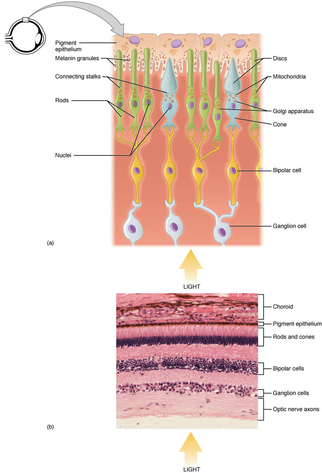
Inner segment The inner segment contains the nucleus and essential organelles, supporting the metabolic needs of photoreceptors. It connects to the outer segment, facilitating energy transfer for visual processing.
Outer segment The outer segment houses membrane arrays with photosensitive opsin molecules, where light absorption occurs. In rods, it forms long columnar shapes with stacked discs containing rhodopsin, while in cones, it features short, tapered shapes with membrane folds.
Rod The rod is a photoreceptor optimized for low-light vision, with a long outer segment filled with rhodopsin pigment. Its high sensitivity makes it ideal for night vision but less effective for color discrimination.
Cone The cone is a photoreceptor specialized for color and high-acuity vision, with a short, tapered outer segment. It contains different opsins sensitive to red, green, or blue light, functioning best in bright conditions.
Nuclei of rods and cones The nuclei of rods and cones form a dense layer in the retina, visible under the microscope at 800x magnification. This layer supports the cellular structure and genetic material needed for photoreceptor function.
Anatomy of Photoreceptors
Photoreceptors are the foundation of the retina’s light-sensing capability, divided into distinct segments. This image provides a dual perspective, showing both the anatomical layout and a microscopic view of these cells.
- The inner segment houses mitochondria and other organelles, providing energy for photoreceptor activity.
- The outer segment contains the photopigment-laden membranes, critical for light detection.
- Rods are more numerous, comprising about 120 million per retina, and dominate in dim light.
- Cones are fewer, around 6 million, and are concentrated in the fovea for detailed vision.
- The nuclei of rods and cones are packed tightly, forming the outer nuclear layer of the retina.
- Supporting cells, like Müller cells, nourish and stabilize these photoreceptors.
- The microscope view at 800x, courtesy of the Regents of University of Michigan Medical School, reveals this cellular density.
Physiology of Photoreceptor Function
Photoreceptors transform light into neural signals through a complex biochemical process. The structural differences between rods and cones underpin their specialized roles in vision.
- The outer segment of rods contains stacked discs, maximizing rhodopsin exposure to light.
- Cones have folded membranes, allowing faster response times for color detection.
- When light hits the outer segment, opsin molecules trigger a cascade, hyperpolarizing the cell.
- Rods are highly sensitive, detecting single photons, but saturate in bright light.
- Cones require more light but provide sharp, color vision via three opsin types.
- The inner segment supports this process by supplying ATP for continuous operation.
- Synaptic connections transmit signals to bipolar and ganglion cells for further processing.
Role of Rods in Low-Light Vision
Rods excel in dim environments, relying on their unique structure for sensitivity. Their design makes them indispensable for night vision and peripheral sight.
- The long outer segment of rods increases surface area for rhodopsin, enhancing light capture.
- Rhodopsin, a G-protein-coupled receptor, breaks down into retinal and opsin upon light absorption.
- This breakdown triggers a signal, amplified through the phototransduction cascade.
- Rods lack color sensitivity, relying on rod-specific bipolar cells for signal relay.
- Their density decreases toward the fovea, explaining reduced acuity in low light.
- Adaptation to darkness involves rhodopsin regeneration, taking 20-30 minutes.
Role of Cones in Color and Detail Vision
Cones are tailored for bright light, with structures optimized for color and acuity. Their distribution and function are key to daytime and detailed visual tasks.
- The tapered outer segment of cones contains folded membranes, speeding up photopigment renewal.
- Three cone types—L, M, and S—detect red, green, and blue light, respectively.
- This trichromatic system allows color perception through opponent processing in the brain.
- Cones are densely packed in the fovea, providing the highest visual resolution.
- Their lower sensitivity requires brighter conditions, complementing rod function.
- Damage to cones can lead to color blindness or reduced central vision.
Microscopic Insights from Retinal Tissue
The microscope view at 800x magnification reveals the cellular architecture of the retina. This high-resolution image, provided by the Regents of University of Michigan Medical School, showcases photoreceptor nuclei.
- The nuclei of rods and cones appear as a thick band, indicating their abundance.
- Rod nuclei outnumber cone nuclei, reflecting their prevalence in the retina.
- The image highlights the outer nuclear layer, where these nuclei reside.
- Adjacent layers, like the outer plexiform layer, contain synaptic connections.
- Staining techniques enhance visibility of these nuclei, aiding histological analysis.
- This view supports studies on retinal health and photoreceptor distribution.
Clinical Relevance of Photoreceptors
Understanding photoreceptor anatomy aids in diagnosing and managing vision disorders. This image provides a baseline for assessing normal retinal structure.
- Retinitis pigmentosa affects rods first, leading to night blindness and tunnel vision.
- Age-related macular degeneration impacts cones in the fovea, causing central vision loss.
- The outer segment damage can result from oxidative stress, common in retinal diseases.
- Cone dysfunction may lead to color vision deficiencies, like red-green color blindness.
- The nuclei of rods and cones layer thinning is a marker in degenerative conditions.
- Treatments like gene therapy target photoreceptor preservation in early stages.
- Regular retinal imaging monitors these structures for timely intervention.
In conclusion, the photoreceptors of the eye, as depicted in this anatomical and microscopic view, are a testament to the complexity of vision. Their specialized structures and functions enable us to navigate diverse lighting conditions, making the retina a fascinating area for exploring ocular health.

