The extraocular muscles are essential components of the eye, enabling precise movements and maintaining alignment within the orbit. This detailed diagram showcases the lateral and anterior perspectives of the right eye, highlighting the muscles and structures that coordinate vision and eye position.
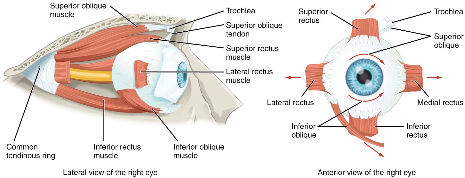
Superior oblique muscle The superior oblique muscle rotates the eye downward and outward, aiding in downward gaze and intorsion. It passes through the trochlea, a pulley-like structure, to adjust its angle of pull.
Trochlea The trochlea is a cartilaginous loop that guides the superior oblique tendon, enhancing its mechanical advantage. This structure ensures smooth movement and precise control during eye rotation.
Superior oblique tendon The superior oblique tendon connects the superior oblique muscle to the eyeball, transmitting force through the trochlea. It allows the muscle to exert its effect at an optimal angle for complex eye movements.
Superior rectus muscle The superior rectus muscle elevates the eye and assists in inward rotation, contributing to upward gaze. It is innervated by the oculomotor nerve, ensuring coordinated action with other muscles.
Lateral rectus muscle The lateral rectus muscle abducts the eye, moving it outward away from the nose. This muscle is controlled by the abducens nerve, providing lateral movement essential for tracking objects.
Inferior rectus muscle The inferior rectus muscle depresses the eye and aids in outward rotation, facilitating downward gaze. It works in conjunction with the superior rectus for vertical eye alignment.
Inferior oblique muscle The inferior oblique muscle elevates and outwardly rotates the eye, supporting upward and lateral movements. Its unique insertion pattern enhances its role in complex eye tracking.
Medial rectus muscle The medial rectus muscle adducts the eye, moving it inward toward the nose. Innervated by the oculomotor nerve, it balances the lateral rectus for horizontal eye alignment.
Common tendinous ring The common tendinous ring is a fibrous band where the four rectus muscles originate, anchoring them to the orbit. This structure provides a stable base for coordinated eye movements.
Anatomy of the Extraocular Muscles
The extraocular muscles surround the eyeball, working in harmony to control its position and movement. These muscles, depicted in lateral and anterior views, are crucial for maintaining visual focus and alignment.
- The six extraocular muscles include four rectus and two oblique muscles, each with distinct actions.
- The orbit, a bony cavity, houses these muscles, protecting the eye while allowing mobility.
- The superior and inferior rectus muscles primarily handle vertical movements, with secondary rotational effects.
- The lateral and medial rectus muscles govern horizontal eye motion, ensuring binocular vision.
- The oblique muscles, superior and inferior, add torsional and diagonal capabilities to eye movement.
- Tendons, like the superior oblique tendon, transmit muscle force to the eyeball’s surface.
- The common tendinous ring, or annulus of Zinn, serves as the origin point for the rectus muscles.
Functions of Eye Movement
Each extraocular muscle contributes to specific eye movements, enabling a wide range of visual tasks. Their coordinated action ensures smooth tracking and stable gaze.
- The superior rectus muscle lifts the eye, assisting in looking up or reading overhead signs.
- The lateral rectus muscle moves the eye outward, crucial for peripheral vision or following moving objects.
- The inferior rectus muscle lowers the eye, aiding in downward gaze like reading or inspecting the floor.
- The medial rectus muscle brings the eye inward, essential for focusing on close objects.
- The superior oblique muscle, via the trochlea, rotates the eye downward and outward for precise adjustments.
- The inferior oblique muscle elevates and rotates the eye, supporting upward and lateral tracking.
- These muscles work in pairs, with agonist and antagonist actions for balanced movement.
- Rapid eye movements, or saccades, rely on this muscular synergy for quick shifts in focus.
Role of the Trochlea and Tendons
The trochlea and associated tendons enhance the mechanical efficiency of the superior oblique muscle. This setup allows for intricate control over eye rotation.
- The trochlea acts as a pulley, redirecting the superior oblique tendon to optimize its pull angle.
- The superior oblique tendon, passing through the trochlea, reduces friction and wear during movement.
- This configuration enables the muscle to exert torque, rotating the eye along its visual axis.
- Tendons are composed of dense collagen, providing strength and flexibility.
- The trochlea’s cartilage composition minimizes stress on the tendon during repeated use.
- Misalignment of the trochlea, though rare, can affect superior oblique function, impacting gaze.
Innervation and Coordination
The extraocular muscles are innervated by cranial nerves, ensuring precise and rapid responses. This neural control integrates with the brain for seamless eye movement.
- The oculomotor nerve (CN III) supplies the superior, inferior, and medial rectus muscles.
- The trochlear nerve (CN IV) innervates the superior oblique muscle, guiding its unique path.
- The abducens nerve (CN VI) controls the lateral rectus muscle, specializing in abduction.
- These nerves originate from the brainstem, with nuclei coordinating bilateral eye movements.
- Reflex arcs, like the vestibulo-ocular reflex, stabilize gaze during head motion.
- Damage to these nerves can lead to strabismus or diplopia, though this diagram shows normal anatomy.
Clinical Relevance of Extraocular Muscles
Understanding these muscles aids in diagnosing and managing eye movement disorders, though this image depicts healthy structures. Knowledge of their anatomy supports effective treatment strategies.
- Weakness in the lateral rectus muscle may cause esotropia, where the eye turns inward.
- Superior oblique dysfunction, linked to trochlea issues, can result in cyclotorsion or vertical diplopia.
- The inferior rectus muscle’s impairment might lead to difficulty with downward gaze.
- Medial rectus overaction can contribute to convergence insufficiency, affecting near vision.
- Muscle balance is assessed via the cover test or eye movement recordings.
- Surgical correction, such as recession or resection, adjusts muscle tension in strabismus cases.
- Rehabilitation exercises strengthen these muscles in cases of nerve palsy.
In conclusion, the extraocular muscles of the eye, as illustrated in this diagram, form a sophisticated system for movement and stability. Their precise coordination, supported by tendons and neural innervation, underscores the complexity of visual function and its maintenance.

