The human eye, nestled within the protective orbit of the skull, is a complex organ supported by surrounding tissues that ensure its functionality and safety. This detailed view highlights the intricate anatomy, including muscles, membranes, and protective features that work together to maintain vision and ocular health.
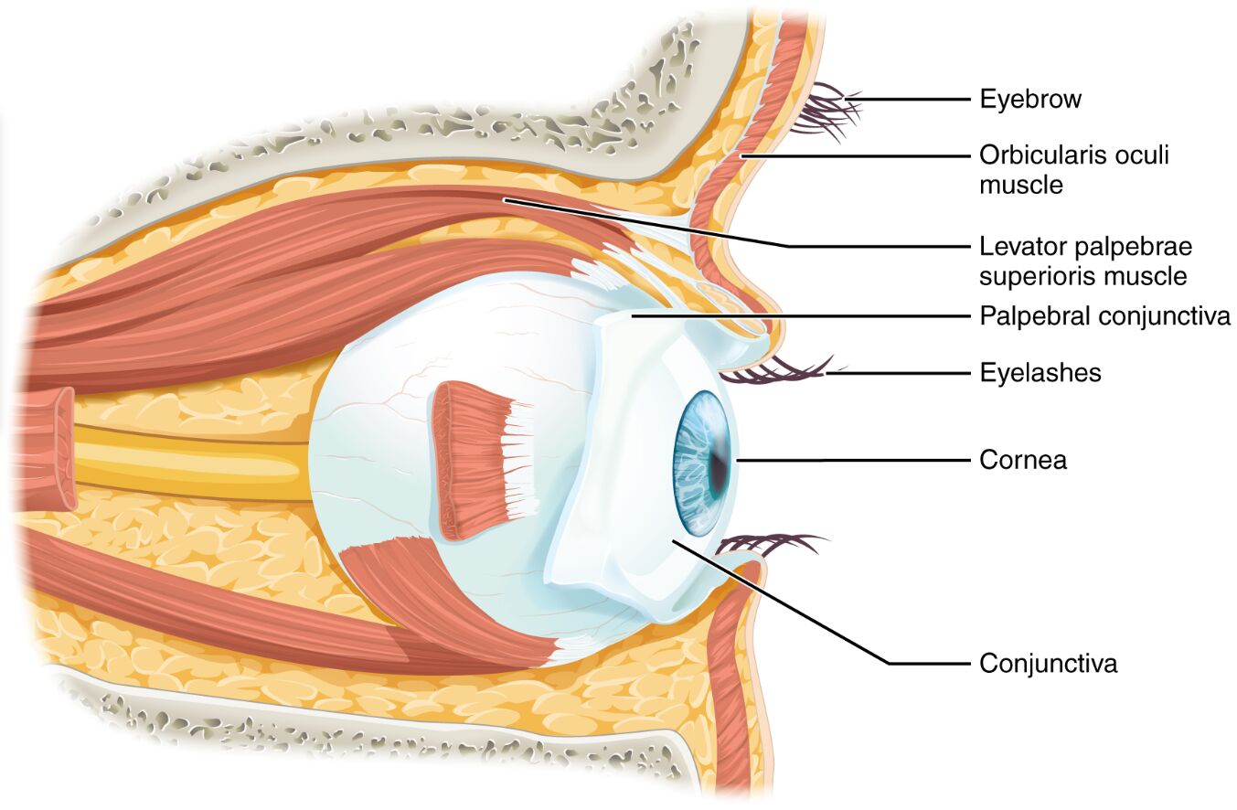
Eyebrow The eyebrow serves as a protective barrier, shielding the eye from sweat, debris, and sunlight. It also plays a role in facial expression and helps channel moisture away from the eye.
Orbicularis oculi muscle The orbicularis oculi muscle is a circular muscle around the eye that facilitates blinking and eyelid closure. This action protects the cornea and spreads tears to maintain moisture.
Levator palpebrae superioris muscle The levator palpebrae superioris muscle elevates the upper eyelid, enabling the eye to open and adjust to light. It is innervated by the oculomotor nerve, ensuring precise control of eyelid movement.
Palpebral conjunctiva The palpebral conjunctiva is a thin, moist membrane lining the inner surface of the eyelids. It protects the eye by preventing friction and supporting tear distribution.
Eyelashes The eyelashes act as a first line of defense, trapping dust and small particles before they reach the eye. They also contribute to sensory feedback by detecting nearby objects.
Cornea The cornea is the transparent front layer of the eye that refracts light to focus it onto the retina. It is avascular, relying on diffusion from tears and aqueous humor for nourishment.
Conjunctiva The conjunctiva is a thin mucous membrane covering the front of the eye and lining the eyelids. It provides lubrication and protection while allowing smooth movement of the eyeball.
Overview of Eye Anatomy
The eye resides within the orbit, a bony cavity formed by cranial bones that offer robust protection. Surrounding soft tissues and muscles enhance its functionality and safeguard its delicate structures.
- The orbit, composed of seven bones including the frontal and zygomatic, creates a secure housing for the eye.
- Adipose tissue within the orbit cushions the eye, absorbing shocks and maintaining position.
- The extraocular muscles, such as the levator palpebrae superioris, control eye and eyelid movement.
- The conjunctiva extends from the cornea to the inner eyelids, ensuring a moist environment.
- Blood vessels and nerves, like those innervating the orbicularis oculi, support metabolic needs and motor control.
- The cornea and sclera form the outer coat, with the cornea being the primary refractive surface.
Functions of Orbital Structures
Each component of the eye and orbit contributes to vision and protection through specialized roles. The interplay of muscles, membranes, and external features maintains optimal eye health.
- The eyebrow deflects sweat and debris, reducing the risk of irritation or infection.
- The orbicularis oculi muscle contracts during blinking, spreading the tear film across the cornea.
- The levator palpebrae superioris muscle lifts the eyelid, allowing light to enter and vision to occur.
- The palpebral conjunctiva secretes mucus to lubricate the eye, preventing dryness.
- Eyelashes trigger a reflex blink when touched, offering an immediate protective response.
- The cornea focuses incoming light, with its curvature adjusted by tear film thickness.
- The conjunctiva filters out minor particles, supporting the eye’s immune defense.
Muscular Support and Movement
Muscles around the eye enable precise movements and protective actions essential for vision. Their coordinated function ensures the eye adapts to various conditions.
- The orbicularis oculi muscle is divided into orbital and palpebral parts, each serving distinct closure strengths.
- The levator palpebrae superioris muscle works with Müller’s muscle for fine-tuned elevation.
- These muscles are controlled by cranial nerves III, V, and VII, ensuring rapid responses.
- Contraction patterns vary, with voluntary blinks lasting about 0.3 seconds.
- Fatigue or nerve damage can impair muscle function, affecting eyelid control.
- Regular exercise of these muscles supports long-term ocular health.
Protective Layers and Surfaces
The eye’s external layers and appendages provide a barrier against environmental hazards. These structures maintain clarity and integrity of the visual system.
- The eyebrow hair growth pattern directs moisture laterally, away from the eye.
- Eyelashes are embedded with sebaceous glands, releasing oils to prevent tear evaporation.
- The cornea’s avascular nature avoids blood vessel interference with light transmission.
- The conjunctiva contains goblet cells that produce mucin for tear stability.
- The palpebral conjunctiva reflects onto the bulbar conjunctiva, forming a continuous shield.
- Minor abrasions heal quickly due to the cornea’s regenerative epithelial layer.
- Inflammation of the conjunctiva, or conjunctivitis, highlights its protective role.
Clinical Relevance of Eye Anatomy
Understanding the eye’s anatomy aids in diagnosing and treating ocular conditions, though this image depicts normal structure. Knowledge of these components guides clinical interventions.
- The orbicularis oculi muscle’s dysfunction can lead to lagophthalmos, where the eye fails to close fully.
- The levator palpebrae superioris muscle’s weakness may cause ptosis, or drooping eyelids.
- Corneal abrasions require prompt treatment to prevent infection due to its exposure.
- Conjunctivitis, an inflammation of the conjunctiva, can be bacterial or viral, treated accordingly.
- Eyelash disorders like trichiasis involve inward growth, irritating the cornea.
- Regular eye exams assess the integrity of these structures for early detection of issues.
- Surgical techniques, such as blepharoplasty, may address excess skin affecting the levator muscle.
In summary, the eye within the orbit is a marvel of anatomical design, supported by muscles, membranes, and protective features. This intricate system ensures clear vision and resilience, making it a fascinating subject for exploring human physiology.

