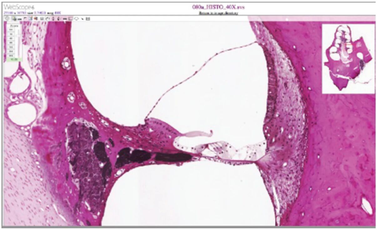
The cochlea and its intricate organ of Corti, captured at a magnification of 412x, reveal the microscopic wonders that underpin human hearing within the inner ear. This image showcases the delicate structures responsible for converting sound vibrations into electrical signals, offering a glimpse into the organ of Corti’s hair cells and their surrounding environment. This article explores the anatomical details and physiological roles of these components, providing a comprehensive understanding of their contribution to auditory perception.

Labeled Parts of the Cochlea and Organ of Corti
Cochlea The cochlea is a spiral, fluid-filled structure in the inner ear that houses the organ of Corti and processes sound vibrations into neural signals. Its coiled design maximizes space efficiency while allowing for frequency-specific sound detection along its length.
Organ of Corti The organ of Corti is a specialized structure within the cochlear duct, resting on the basilar membrane, and contains hair cells that detect sound. It serves as the primary site of auditory transduction, converting mechanical energy into electrical impulses for the brain.
Hair cells Hair cells are mechanoreceptor cells within the organ of Corti, featuring stereocilia that bend with fluid motion to generate electrical signals. They are critical for translating sound vibrations into neural activity, with inner and outer types performing distinct roles.
Basilar membrane The basilar membrane supports the organ of Corti and varies in width and stiffness along the cochlea, tuning it to different sound frequencies. Its movement in response to fluid pressure waves activates specific hair cells for pitch perception.
Tectorial membrane The tectorial membrane is a gelatinous structure overlying the hair cells, interacting with their stereocilia during sound-induced fluid motion. It enhances the mechanical stimulation of hair cells, amplifying the auditory signal.
Stereocilia Stereocilia are hair-like projections on the apical surface of hair cells, bending in response to sound vibrations to open ion channels. This movement triggers the release of neurotransmitters, initiating the auditory signal to the brain.
Anatomical Overview of the Cochlea and Organ of Corti
The microscopic view of the cochlea and organ of Corti reveals a highly organized structure designed for auditory processing. This magnification highlights the interplay between fluid dynamics and cellular components.
- Spiral architecture: The cochlea’s spiral shape encloses the organ of Corti, optimizing the detection of a wide range of sound frequencies.
- Sensory hub: The organ of Corti, nestled within the cochlear duct, contains hair cells that are the cornerstone of sound transduction.
- Membrane support: The basilar membrane provides a dynamic base, while the tectorial membrane enhances hair cell stimulation.
- Cellular precision: Stereocilia on hair cells are arranged in precise rows, ensuring accurate detection of fluid movements.
- Fluid environment: The cochlear fluid surrounds these structures, transmitting pressure waves that drive the auditory process.
Physiological Functions of the Organ of Corti
The organ of Corti, with its hair cells, is the epicenter of auditory transduction, converting mechanical sound waves into electrical signals. Its physiological mechanisms are finely tuned to capture and process sound.
- Vibration detection: Hair cells’ stereocilia bend when the basilar membrane moves, triggered by pressure waves in the cochlear fluid.
- Signal generation: This bending opens ion channels, depolarizing hair cells and releasing neurotransmitters to activate auditory nerve fibers.
- Frequency tuning: The basilar membrane’s gradient allows different regions to resonate with specific frequencies, engaging corresponding hair cells.
- Amplification: The tectorial membrane’s interaction with stereocilia amplifies the mechanical input, enhancing sound sensitivity.
- Neural transmission: Electrical impulses from hair cells are relayed to the cochlear nerve, enabling the brain to interpret sound.
Developmental and Cellular Dynamics
The cochlea and organ of Corti develop during embryogenesis, maturing to support hearing by early childhood. This process involves intricate cellular differentiation and structural growth.
- Embryonic formation: The cochlea arises from the otic vesicle, with the organ of Corti forming within the cochlear duct during fetal development.
- Hair cell differentiation: Hair cells develop with stereocilia, aligning to detect fluid motion as the cochlea matures.
- Membrane development: The basilar membrane grows with a stiffness gradient, enabling frequency-specific responses along its length.
- Tectorial integration: The tectorial membrane forms above the hair cells, establishing its role in sound amplification during prenatal stages.
- Postnatal refinement: Neural connections with hair cells strengthen postnatally, enhancing the cochlea’s auditory sensitivity.
Clinical Relevance and Cochlear Health
Understanding the microscopic structure of the cochlea and organ of Corti is essential for diagnosing and managing hearing impairments. Clinical evaluations often focus on these components to assess auditory function.
- Sensorineural hearing loss: Damage to hair cells, often from noise exposure or ototoxic drugs, disrupts sound transduction in the organ of Corti.
- Presbycusis: Age-related degeneration of hair cells and the basilar membrane leads to high-frequency hearing loss.
- Tinnitus: Persistent ringing may arise from hair cell dysfunction or cochlear damage, impacting quality of life.
- Diagnostic tools: Otoacoustic emission tests and cochlear microscopy evaluate hair cell health and membrane integrity.
- Therapeutic options: Cochlear implants replace damaged hair cells, while protective measures like noise reduction preserve remaining function.
In conclusion, the microscopic view of the cochlea and organ of Corti, as captured in this image, unveils the intricate machinery behind hearing, with hair cells and their supporting membranes at the forefront. The interplay of the basilar and tectorial membranes with stereocilia highlights the precision required to transform sound into perception, offering a window into auditory health. Exploring these structures enhances appreciation for the cochlea’s role and informs strategies to maintain its functionality.

