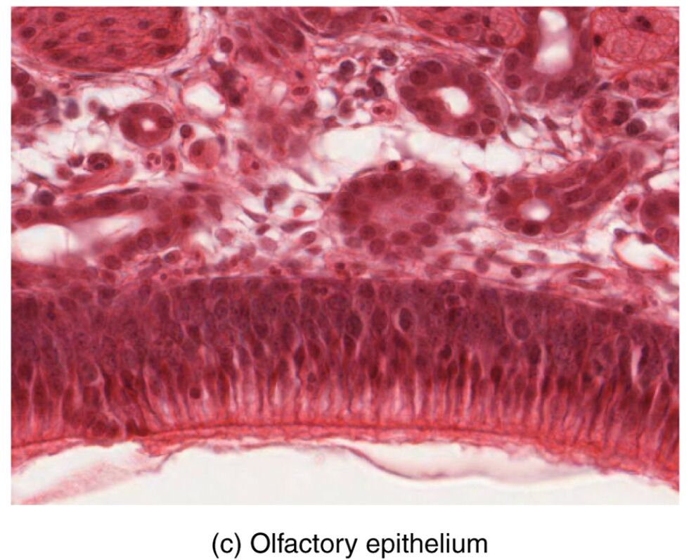
The olfactory epithelium, a vital component of the olfactory system, reveals its intricate cellular architecture when viewed under a microscope, offering a glimpse into the foundation of smell perception. This image, captured at a magnification of 812x, illustrates how axons from olfactory receptor neurons extend through the cribriform plate to synapse with neurons in the olfactory bulb, highlighting the system’s connectivity. This article explores the microscopic structure and physiological significance of the olfactory epithelium, providing a thorough understanding of its role in sensory processing.
Labeled Parts of the Olfactory Epithelium
Olfactory epithelium The olfactory epithelium is a specialized pseudostratified tissue lining the nasal cavity, housing olfactory receptor neurons that detect odorant molecules. It is supported by a mucus layer and contains supporting cells, ensuring a functional sensory surface.
Olfactory receptor neuron The olfactory receptor neuron is a bipolar cell within the olfactory epithelium, with dendrites bearing cilia that detect odors and an axon that transmits signals. Its axons bundle to form the olfactory nerve, connecting the nasal cavity to the brain.
Axon The axon is the long process of an olfactory receptor neuron that carries electrical impulses from the cell body to the olfactory bulb. These axons penetrate the cribriform plate, synapsing with bulb neurons to relay smell information.
Cribriform plate The cribriform plate is a perforated section of the ethmoid bone that allows axons of olfactory receptor neurons to pass from the nasal cavity to the olfactory bulb. It serves as a protective yet permeable barrier, facilitating neural connectivity.
Olfactory bulb neuron The olfactory bulb neuron, typically a mitral or tufted cell, receives synaptic input from olfactory receptor neuron axons within the olfactory bulb. It processes and integrates odor signals, transmitting them to higher brain regions via the olfactory tract.
Supporting cell The supporting cell, or sustentacular cell, provides structural and metabolic support to olfactory receptor neurons within the epithelium. It also contributes to the mucus layer and detoxifies harmful substances, protecting the sensory cells.
Mucus layer The mucus layer covers the olfactory epithelium, dissolving odorant molecules to enable detection by receptor neurons. It is secreted by underlying glands, maintaining a moist environment essential for olfactory function.
Cilia Cilia are microscopic hair-like projections on the dendrites of olfactory receptor neurons, increasing the surface area for odorant binding. They move within the mucus, enhancing the efficiency of smell detection.
Anatomical Overview of the Olfactory Epithelium
The olfactory epithelium, as seen under the microscope, showcases a complex arrangement of cells and structures critical to olfaction. This high-magnification view reveals the interplay between sensory and supportive elements.
- Cellular layers: The epithelium includes olfactory receptor neurons, supporting cells, and basal cells, forming a dynamic sensory tissue.
- Axonal pathway: Axons extend from receptor neurons, passing through the cribriform plate to reach the olfactory bulb, establishing the neural link.
- Protective coating: The mucus layer traps odorants and shields the epithelium, while cilia on neuron dendrites maximize exposure to these molecules.
- Structural support: Supporting cells maintain the integrity of the epithelium, providing nutrients and detoxification capabilities.
- Bone interface: The cribriform plate’s perforations allow precise axonal passage, balancing protection with connectivity to the brain.
Physiological Functions of the Olfactory Epithelium
The olfactory epithelium converts chemical stimuli into neural signals, forming the first step in smell perception. Its microscopic features enable efficient detection and transmission of odor information.
- Odorant detection: Cilia on olfactory receptor neurons bind to odorant molecules dissolved in the mucus, initiating a sensory response.
- Signal generation: This binding triggers a G-protein-coupled cascade, producing electrical impulses that travel along the axon.
- Neural relay: Axons project through the cribriform plate, synapsing with olfactory bulb neurons to process and refine the smell signal.
- Supportive role: Supporting cells nourish receptor neurons and maintain the mucus layer, ensuring optimal sensory conditions.
- Adaptive response: The epithelium adapts to continuous odor exposure, with receptor sensitivity adjusting to focus on new stimuli.
Developmental and Cellular Dynamics
The olfactory epithelium develops during embryogenesis, with its cellular components maturing to support smell by birth. Ongoing regeneration ensures its functionality over time.
- Embryonic formation: The epithelium arises from ectodermal tissue, with receptor neurons and supporting cells differentiating early in gestation.
- Cellular renewal: Basal cells, not visible in this micrograph, regenerate receptor neurons every few weeks, maintaining sensory capacity.
- Axonal growth: Axons extend through the cribriform plate during development, forming synapses with bulb neurons prenatally.
- Ciliary development: Cilia form on neuron dendrites, enhancing detection as the system matures postnatally.
- Evolutionary adaptation: This regenerative capacity reflects an evolutionary trait to sustain olfaction amidst environmental challenges.
Clinical Relevance and Olfactory Considerations
The microscopic structure of the olfactory epithelium is key to diagnosing and managing smell-related conditions. Clinical evaluations often focus on this tissue to detect abnormalities.
- Anosmia: Loss of smell can result from damage to olfactory receptor neurons or their axons, often due to head trauma or infections.
- Hyposmia: Reduced smell sensitivity may indicate epithelial degeneration or axonal impairment, commonly linked to aging or disease.
- Diagnostic tools: Microscopic analysis and olfactory testing with odorants assess epithelial health and neural function.
- Therapeutic strategies: Treatments include addressing inflammation, supporting regeneration with vitamins, or surgical intervention for cribriform plate issues.
- Health implications: Impaired olfaction can affect taste, nutrition, and safety, highlighting the need for early detection and management.
In conclusion, the olfactory epithelium, as revealed under the microscope, exemplifies the intricate design of the olfactory system, from receptor neurons to axonal projections. This microscopic view underscores the epithelium’s role in detecting odors and transmitting signals to the brain, offering valuable insights into sensory physiology and potential clinical applications. Understanding these structures enhances our ability to address olfactory health and maintain overall sensory well-being.

