The olfactory system, a cornerstone of the human sensory network, initiates its remarkable process within the nasal cavity, where it detects and interprets a vast array of odors. This image illustrates the key structures involved, highlighting the pathway of inhaled air and the connection to the brain, which together enable the perception of smell. This article provides a detailed exploration of these anatomical features, offering insights into their roles and significance in olfactory function.
Labeled Parts of the Olfactory System
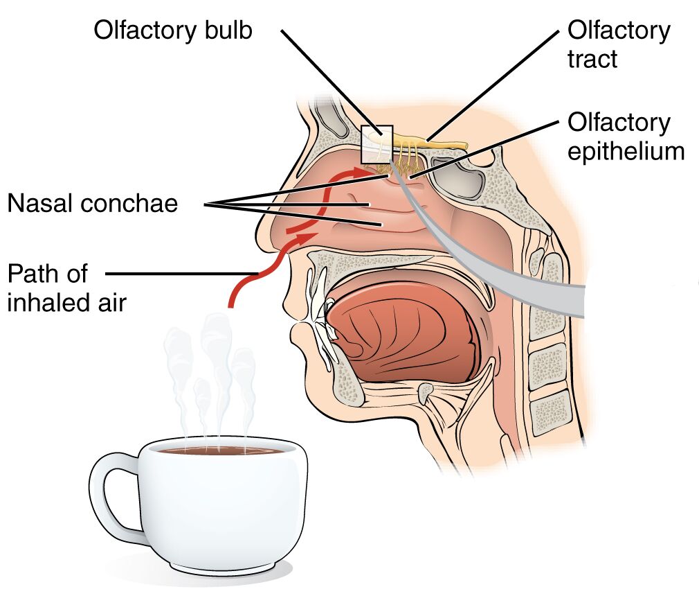
Olfactory bulb The olfactory bulb is a neural structure at the base of the brain that receives signals from olfactory neurons via their axons. It processes these signals and relays them to other brain regions through the olfactory tract, playing a pivotal role in smell perception.
Olfactory tract The olfactory tract is a bundle of nerve fibers extending from the olfactory bulb to the olfactory cortex, facilitating the transmission of processed odor information. It serves as a critical link between the peripheral olfactory system and higher brain centers for conscious smell recognition.
Olfactory epithelium The olfactory epithelium is a specialized tissue lining the upper nasal cavity, containing olfactory receptor neurons that detect odorant molecules. It acts as the primary sensory interface, converting chemical stimuli into neural signals.
Nasal conchae The nasal conchae are bony projections within the nasal cavity that increase air turbulence and surface area. They direct inhaled air toward the olfactory epithelium, enhancing the efficiency of odorant molecule detection.
Path of inhaled air The path of inhaled air travels through the nasal cavity, guided by the nasal conchae to reach the olfactory epithelium. This airflow carries odorant molecules, initiating the olfactory process by bringing them into contact with receptor cells.
Anatomical Overview of the Olfactory System
The olfactory system’s foundation lies in the nasal cavity, where anatomical structures work in harmony to capture and process odors. This intricate design ensures that even subtle scents are effectively detected and transmitted.
- Airflow dynamics: The nasal conchae create turbulence, ensuring that inhaled air rich with odorants contacts the olfactory epithelium efficiently.
- Epithelial role: The olfactory epithelium houses receptor cells embedded in a moist environment, ready to interact with airborne molecules.
- Neural connection: Axons from the olfactory epithelium extend to the olfactory bulb, forming the initial segment of the olfactory pathway.
- Brain integration: The olfactory tract carries processed signals from the bulb to the brain, enabling the perception and analysis of smells.
- Protective features: The nasal cavity’s structure filters and humidifies air, safeguarding the delicate olfactory epithelium while supporting its function.
Physiological Functions of the Olfactory System
The olfactory system transforms chemical cues in the air into sensory experiences, playing an essential role in daily life. Its physiological mechanisms are finely tuned to detect and interpret a wide range of odors.
- Odorant detection: Olfactory receptors within the epithelium bind to odorant molecules carried by inhaled air, triggering a neural response.
- Signal initiation: The olfactory epithelium converts chemical stimuli into electrical impulses, which are transmitted via neurons to the brain.
- Signal processing: The olfactory bulb refines these impulses, with mitral cells enhancing the signal before it travels along the olfactory tract.
- Sensory integration: The brain interprets these signals, linking them to memory, emotion, and even taste perception for a holistic sensory experience.
- Adaptive response: The system adjusts to continuous exposure, allowing it to distinguish new odors amidst background scents.
Developmental and Cellular Dynamics
The olfactory system develops early in fetal life, with its structures maturing to support smell perception shortly after birth. Ongoing cellular processes ensure its functionality throughout life.
- Embryonic development: The olfactory epithelium forms from ectodermal tissue, with receptor neurons differentiating during gestation.
- Cellular turnover: Olfactory neurons are regularly replaced by stem cells within the epithelium, maintaining sensory capability over time.
- Neural growth: Axons from the epithelium extend to the olfactory bulb, establishing connections that refine during early development.
- Structural evolution: The nasal conchae develop to optimize airflow, reflecting an evolutionary adaptation for enhanced olfaction.
- Age-related changes: Sensory decline may occur with age, potentially due to reduced epithelial cell efficiency or neuronal loss.
Clinical Relevance and Olfactory Considerations
Understanding the olfactory system’s anatomy is key to recognizing and addressing smell-related issues. Clinical evaluations often focus on these structures to diagnose potential dysfunctions.
- Anosmia: Complete loss of smell can result from damage to the olfactory epithelium or bulb, often due to head trauma or infections.
- Hyposmia: Reduced smell sensitivity may indicate aging or neurological conditions, impacting safety and nutrition.
- Diagnostic approaches: Olfactory testing with specific odors and imaging of the nasal cavity assess system integrity.
- Therapeutic strategies: Treatments may include addressing underlying causes like sinus issues or using olfactory training to stimulate recovery.
- Associated impacts: Loss of smell can affect appetite and hazard detection, emphasizing the need for timely intervention.
In conclusion, the olfactory system’s anatomy within the nasal cavity represents a fascinating interplay of structure and function, enabling the rich sensory experience of smell. From the airflow guided by nasal conchae to the neural processing in the olfactory bulb, this system underscores the body’s ability to adapt and respond to its environment, offering valuable insights for both health and research.

