The tongue serves as a critical organ for taste perception, featuring a complex network of papillae and taste buds that detect chemical compounds in food and drink. These structures, illustrated in this image, are integral to the sensory experience, connecting to the facial and glossopharyngeal nerves to relay taste information to the brain. This article delves into the anatomical details and physiological roles of these components, offering a comprehensive look at how the tongue processes one of our primary senses.
Labeled Parts of the Tongue’s Nerve Structure
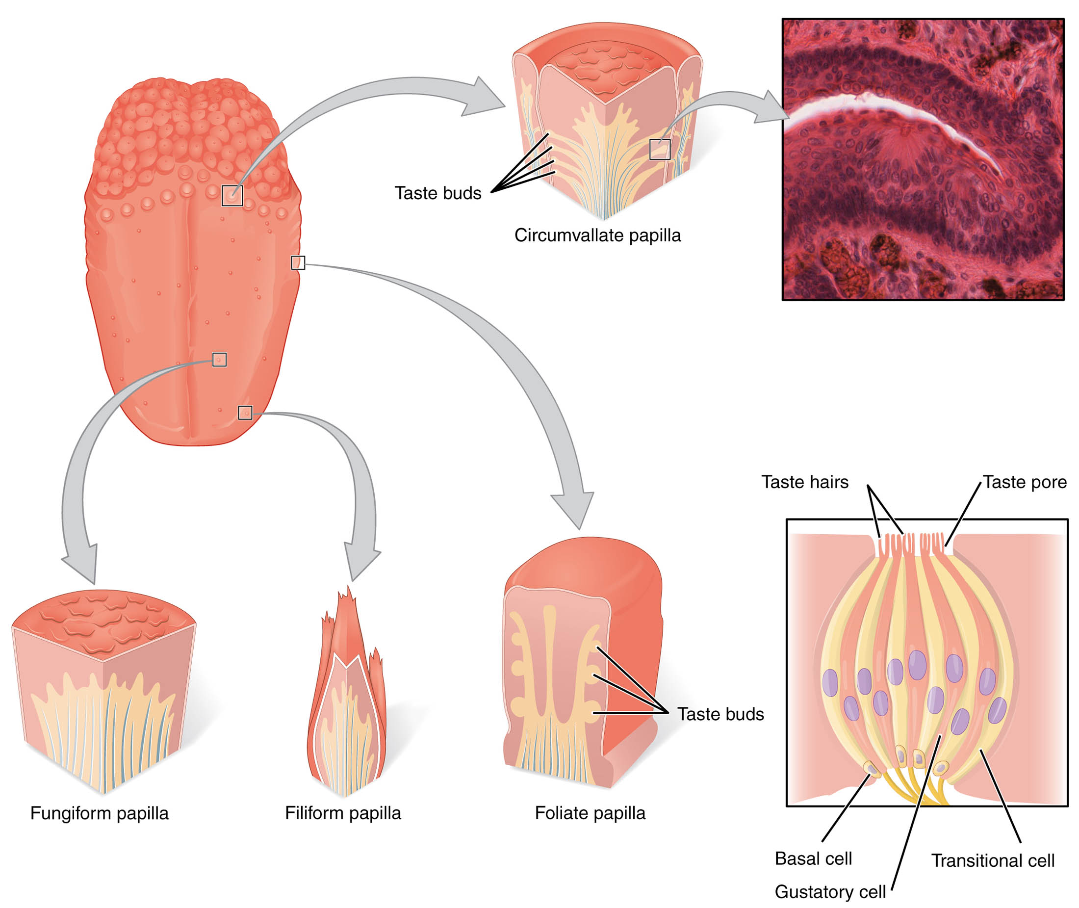
Taste buds Taste buds are microscopic sensory organs embedded within the papillae of the tongue, housing gustatory receptor cells that detect sweet, sour, salty, bitter, and umami tastes. They are constantly renewed every 10-14 days, ensuring the tongue maintains its sensitivity to flavor changes throughout life.
Circumvallate papilla The circumvallate papilla is a large, dome-shaped structure located at the back of the tongue, surrounded by a trench and containing numerous taste buds. These papillae are particularly sensitive to bitter tastes, playing a key role in detecting potentially harmful substances in food.
Filiform papilla The filiform papilla is a conical, thread-like structure covering most of the tongue’s surface, providing texture and aiding in food manipulation. Unlike other papillae, it lacks taste buds but contributes to the tongue’s rough surface, enhancing grip during chewing.
Foliate papilla The foliate papilla consists of folds or ridges on the lateral edges of the tongue, housing taste buds that are sensitive to multiple taste modalities. These structures are more prominent in younger individuals and may diminish with age, affecting taste perception.
Taste hairs Taste hairs are microvilli extending from the gustatory cells within taste buds, increasing the surface area for chemical detection. They project into the taste pore, where they interact with dissolved food molecules to initiate the taste signal.
Taste pore The taste pore is a small opening on the surface of a taste bud, allowing saliva and dissolved food particles to reach the taste hairs. This gateway is essential for the transduction of chemical stimuli into electrical signals within the gustatory cells.
Basal cell The basal cell is a stem cell located at the base of the taste bud, responsible for regenerating gustatory and transitional cells. Its continuous division ensures the taste bud’s functional longevity and adaptability to environmental changes.
Transitional cell The transitional cell serves as an intermediate stage between basal cells and mature gustatory cells, supporting the renewal process within the taste bud. It gradually differentiates into specialized cells, contributing to the taste bud’s dynamic structure.
Gustatory cell The gustatory cell is a specialized receptor cell within the taste bud that detects chemical stimuli and transmits taste information to sensory neurons. It contains taste hairs that extend to the taste pore, where they bind with taste molecules to trigger neural signals.
Anatomical Overview of the Tongue’s Nerve Structure
The tongue’s surface is adorned with various papillae, each contributing to its sensory and mechanical functions. This intricate anatomy supports the detection and processing of taste, linking directly to the nervous system.
- Papillae distribution: The tongue features circumvallate, filiform, and foliate papillae, with each type localized to specific regions, optimizing taste and texture perception.
- Taste bud location: Taste buds are primarily housed in circumvallate and foliate papillae, with their density varying across the tongue’s surface for differential taste sensitivity.
- Microscopic features: The taste pore and taste hairs facilitate chemical interaction, while basal and transitional cells ensure cellular turnover within the taste buds.
- Nerve connections: Gustatory cells synapse with sensory neurons from the facial (VII) and glossopharyngeal (IX) nerves, transmitting taste signals to the brainstem.
- Structural support: The filiform papilla, though lacking taste buds, enhances mechanical function, complementing the sensory roles of other papillae.
Physiological Functions of Taste Structures
The tongue’s nerve structure is designed to detect and interpret chemical stimuli, playing a vital role in nutrition and safety. These functions rely on the coordinated activity of taste buds and associated cells.
- Taste detection: Gustatory cells within taste buds respond to five basic tastes—sweet, sour, salty, bitter, and umami—via specific receptor proteins.
- Signal transduction: Taste hairs and the taste pore allow chemical molecules to bind with gustatory cells, generating action potentials that travel via sensory nerves.
- Cellular renewal: Basal cells continuously produce transitional and gustatory cells, maintaining taste sensitivity despite constant exposure to food and wear.
- Protective role: The circumvallate papilla’s sensitivity to bitter tastes helps identify toxic substances, triggering avoidance behaviors.
- Mechanical aid: Filiform papillae assist in food manipulation and swallowing, supporting the tongue’s dual sensory and motor functions.
Developmental and Cellular Dynamics
The tongue’s nerve structures develop early in embryogenesis, with papillae and taste buds forming through complex cellular processes. This development ensures functional maturity by birth, with ongoing regeneration throughout life.
- Embryonic origins: Taste buds arise from epithelial thickening, while papillae form from ectodermal and mesodermal interactions during fetal development.
- Cell differentiation: Basal cells differentiate into gustatory and transitional cells, driven by genetic and environmental cues, maintaining taste bud integrity.
- Nerve integration: Sensory neurons from cranial nerves VII and IX establish synapses with gustatory cells, refining taste pathways during postnatal growth.
- Age-related changes: The density of foliate and circumvallate papillae may decrease with age, potentially altering taste perception in older individuals.
- Evolutionary perspective: The distribution of taste buds reflects an evolutionary adaptation to detect nutrients and toxins, enhancing survival.
Clinical Relevance and Taste Disorders
Understanding the tongue’s nerve structure is crucial for diagnosing and managing taste-related conditions. Clinical evaluations often assess taste bud function to identify underlying issues.
-
- Ageusia and hypogeusia: Loss or reduced taste sensation can result from damage to gustatory cells or cranial nerve dysfunction, often linked to infections or nutritional deficiencies.
- Dysgeusia: Altered taste perception, such as a metallic taste, may stem from taste bud inflammation or nerve irritation, requiring targeted treatment.
- Diagnostic methods: Taste testing with chemical solutions and microscopic examination of papillae help evaluate taste bud health.
- Therapeutic approaches: Vitamin B12 supplementation or nerve stimulation can address taste deficits, depending on the cause.
- Associated conditions: Diseases like Sjögren’s syndrome may affect saliva production, indirectly impacting taste pore function and taste sensitivity.
In conclusion, the tongue’s nerve structure, with its diverse papillae and taste buds, exemplifies the body’s sophisticated sensory apparatus. This intricate system not only enhances our enjoyment of food but also serves as a protective mechanism, with ongoing cellular renewal ensuring its resilience. Exploring these structures provides valuable insights into sensory physiology and potential therapeutic interventions.

