The dorsal root ganglion is a key structure in the peripheral nervous system, housing the cell bodies of sensory neurons that relay critical information from the body to the spinal cord. This photomicrograph provides a detailed view of its cellular organization, showcasing unipolar neurons and their axons, which form part of the dorsal nerve root. Exploring this anatomy offers a deeper understanding of sensory processing and the intricate network that supports bodily sensation.
Labeled Structures in Dorsal Root Ganglion
This section explores each labeled component in the provided image, offering insights into their anatomical roles and significance.
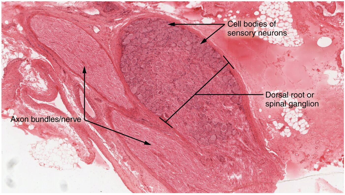
Dorsal root ganglion The dorsal root ganglion is a swelling on the dorsal root of a spinal nerve, containing the cell bodies of unipolar sensory neurons that transmit sensory information like touch and pain. These neurons are clustered within a capsule of connective tissue, providing a protected environment for signal processing.
Unipolar neuron cell body The unipolar neuron cell body is the central part of sensory neurons in the dorsal root ganglion, featuring a single process that splits into peripheral and central branches. This structure allows the neuron to receive sensory input from the periphery and relay it to the spinal cord, optimizing sensory signal transmission.
Axons The axons are the elongated projections extending from the unipolar neuron cell bodies, forming the fibrous region of the dorsal root ganglion. These axons carry sensory impulses toward the spinal cord, bundling together to become part of the dorsal nerve root for further central processing.
Anatomy of the Dorsal Root Ganglion
The dorsal root ganglion’s structure reflects its specialized role in sensory transmission. This anatomical layout ensures efficient communication between the periphery and the central nervous system.
- The ganglion is encased in a connective tissue capsule derived from the epineurium, offering mechanical protection to the neuron cell bodies.
- Unipolar neurons, with their single axon splitting into two branches, are uniquely adapted for sensory functions, receiving input from receptors and sending signals centrally.
- The axons, organized into a fibrous region, exit the ganglion as part of the dorsal root, connecting to the spinal cord’s dorsal horn.
- Surrounding satellite cells provide metabolic support and insulation, enhancing the ganglion’s ability to process sensory data.
Physiological Role in Sensory Processing
The dorsal root ganglion plays a vital role in interpreting and transmitting sensory information. Its physiological functions are essential for maintaining awareness of the body’s internal and external environment.
- Unipolar neuron cell bodies receive sensory input from receptors in the skin, muscles, and organs, converting mechanical, thermal, or chemical stimuli into electrical signals.
- Axons conduct these signals at speeds up to 120 m/s in myelinated fibers, depending on diameter and myelin thickness, ensuring rapid transmission to the spinal cord.
- The ganglion integrates various sensory modalities, including touch, pain, and temperature, before relaying them to higher brain centers via the central nervous system.
- Neurotransmitters like glutamate are released at synaptic junctions, facilitating the transfer of sensory information to second-order neurons.
Clinical Significance and Microscopic Insights
The microscopic view of the dorsal root ganglion provides valuable insights into its structure and clinical relevance. This level of detail aids in diagnosing and managing sensory-related conditions.
- Peripheral neuropathies, such as those caused by diabetes, can damage unipolar neuron cell bodies, leading to numbness or tingling sensations.
- Axon degeneration in the dorsal root ganglion may occur in conditions like herpes zoster, where viral infection affects sensory nerve fibers.
- The 40x magnification in this micrograph highlights cell body clustering, assisting in the identification of pathological changes like inflammation or atrophy.
- Electromyography (EMG) and nerve conduction studies assess ganglion function, guiding treatments for sensory deficits or neuralgia.
Nerve Regeneration and Sensory Recovery
The dorsal root ganglion supports sensory neuron regeneration, a key factor in recovering from nerve injury. This regenerative capacity underscores its adaptability in maintaining sensory function.
- Unipolar neuron cell bodies can regenerate axons if the peripheral branch is damaged, guided by Schwann cells along the original pathway.
- Axons in the fibrous region may regrow at a rate of 1-2 mm per day, depending on the injury’s severity and the presence of growth factors.
- Satellite cells within the ganglion release neurotrophic factors, such as nerve growth factor (NGF), to promote axon elongation and repair.
- Successful regeneration depends on minimizing scar tissue formation, which can impede axonal regrowth and sensory recovery.
In conclusion, the dorsal root ganglion serves as a critical hub for sensory processing, with its unipolar neurons and axons forming a robust network for transmitting bodily sensations. The microscopic insights provided by this photomicrograph enhance our understanding of its anatomy and physiology, supporting the development of targeted interventions for sensory disorders and nerve injuries.

