The meningeal layers surrounding the brain, particularly around the superior sagittal sinus, form a protective and functional barrier critical for cerebral health. These layers, including the dura mater, arachnoid mater, and pia mater, work in harmony to encase the brain, regulate cerebrospinal fluid (CSF) dynamics, and facilitate venous drainage. Exploring their structure and role provides a deeper understanding of intracranial physiology and the vital processes that sustain brain function.
Labeled Structures in Meningeal Layers
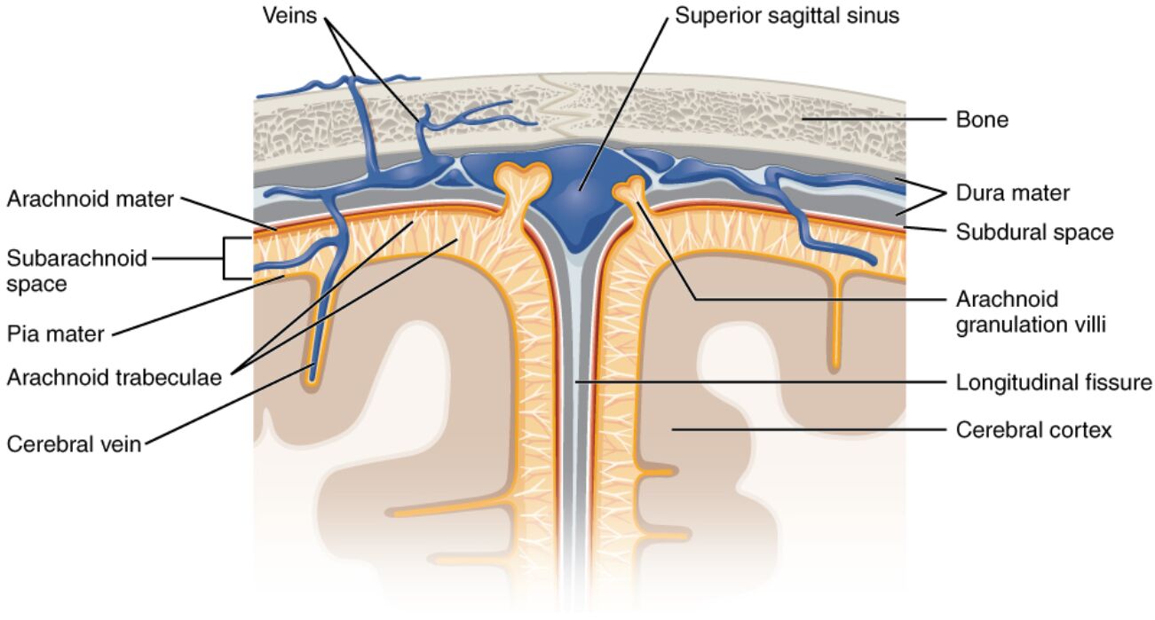
This section provides an overview of each labeled component in the medical image, detailing their anatomical positions and physiological contributions.
Veins The veins are visible as blue structures that drain deoxygenated blood from the cerebral cortex into the superior sagittal sinus, ensuring efficient removal of metabolic waste. These vessels lack valves, relying on the pressure gradient within the sinus to propel blood toward the jugular vein system.
Superior sagittal sinus The superior sagittal sinus is a large venous sinus located along the midline within the dura mater, collecting blood from the cerebral veins and arachnoid granulations. It plays a central role in venous drainage, channeling blood toward the confluence of sinuses and ultimately to the internal jugular vein.
Bone The bone, part of the cranium, forms the rigid outer layer that protects the brain and supports the attachment of the dura mater. It provides a stable framework that houses the meningeal layers and venous sinuses, safeguarding the intracranial contents.
Dura mater The dura mater is the outermost meningeal layer, a tough fibrous tissue adhered to the inner surface of the cranium, enclosing the superior sagittal sinus. It acts as a protective barrier and structural support, with its inner layer reflecting to form the sinus walls.
Subdural space The subdural space is a potential space between the dura mater and arachnoid mater, normally containing a thin film of fluid to reduce friction during brain movement. It can become clinically significant if a subdural hematoma forms due to trauma or ruptured vessels.
Arachnoid mater The arachnoid mater is a delicate, web-like middle meningeal layer situated between the dura and pia mater, containing the subarachnoid space. It supports the arachnoid trabeculae and granulation villi, which are essential for CSF filtration into the venous system.
Subarachnoid space The subarachnoid space lies between the arachnoid and pia mater, filled with CSF that cushions the brain and spinal cord while providing buoyancy. This space contains blood vessels and trabeculae, facilitating nutrient exchange and waste removal.
Pia mater The pia mater is the innermost meningeal layer, closely adhering to the surface of the cerebral cortex, following its contours into the longitudinal fissure. It provides a vascular interface, supporting the delivery of oxygen and nutrients to the brain tissue.
Arachnoid trabeculae The arachnoid trabeculae are delicate fibrous strands extending from the arachnoid mater into the subarachnoid space, connecting it to the pia mater. These structures help maintain the spatial arrangement of the meninges and support CSF circulation.
Cerebral vein The cerebral vein drains blood from the brain parenchyma, merging with the superior sagittal sinus to facilitate venous return. Its superficial location makes it integral to the cortical drainage system, working in tandem with deeper venous networks.
Arachnoid granulation villi The arachnoid granulation villi are protrusions of the arachnoid mater that extend into the superior sagittal sinus, allowing CSF to filter back into the bloodstream. These structures are critical for maintaining intracranial pressure by reabsorbing excess CSF.
Longitudinal fissure The longitudinal fissure is the deep midline groove separating the two cerebral hemispheres, covered by the falx cerebri and containing the superior sagittal sinus. It accommodates the meningeal layers and facilitates the structural division of the brain.
Cerebral cortex The cerebral cortex forms the outer layer of the brain, composed of gray matter responsible for higher functions like thought and sensation. It is enveloped by the pia mater, receiving its blood supply and drainage through the associated venous system.
Anatomy of Meningeal Layers
The meningeal layers provide a multilayered protective and functional system around the brain. Their arrangement ensures both structural integrity and physiological balance within the cranial cavity.
- The dura mater, the toughest layer, consists of two layers: the periosteal layer against the bone and the meningeal layer forming sinus walls.
- The arachnoid mater, with its trabeculae, creates a mesh-like structure that supports the subarachnoid space, filled with CSF produced by the choroid plexus.
- The pia mater, rich in blood vessels, adheres closely to the brain surface, delivering oxygen and nutrients while removing waste.
- The longitudinal fissure houses the falx cerebri, a dural fold that stabilizes the hemispheres and contains the superior sagittal sinus.
Physiological Role in CSF and Venous Dynamics
The meningeal layers and superior sagittal sinus are integral to CSF circulation and venous drainage, maintaining intracranial homeostasis. This system supports brain function by managing fluid and pressure dynamics.
- CSF, produced at a rate of about 0.3-0.4 ml/min, circulates through the subarachnoid space, cushioning the brain against trauma.
- The arachnoid granulation villi reabsorb CSF into the venous blood within the superior sagittal sinus, regulating intracranial pressure between 7-15 mmHg.
- Cerebral veins drain approximately 70% of the brain’s blood volume, with the superior sagittal sinus serving as a primary outflow tract.
- This dynamic interplay prevents conditions like hydrocephalus, where CSF buildup can increase pressure and damage brain tissue.
Clinical Significance and Imaging
The meningeal layers and superior sagittal sinus are clinically relevant due to their involvement in various pathologies. Advanced imaging techniques aid in their assessment and management.
- Subdural hematomas, resulting from trauma, can accumulate in the subdural space, requiring surgical intervention like burr hole drainage.
- Arachnoid granulation villi dysfunction can lead to impaired CSF absorption, contributing to conditions like benign intracranial hypertension.
- Magnetic resonance imaging (MRI) and computed tomography (CT) scans visualize meningeal layers and sinuses, detecting abnormalities such as sinus thrombosis.
- Preventive care focuses on managing head injuries and monitoring for signs of increased intracranial pressure, such as headaches or papilledema.
Blood and CSF Flow Pathways
The flow of blood and CSF through the meningeal layers and superior sagittal sinus ensures efficient waste removal and nutrient supply. This pathway highlights the interconnectedness of cerebral circulation.
- Blood from the cerebral cortex drains into superficial veins, which empty into the superior sagittal sinus along the longitudinal fissure.
- CSF flows from the lateral ventricles through the subarachnoid space, with granulation villi facilitating its return to the venous system.
- The dura mater’s sinus walls guide venous blood toward the confluence of sinuses, maintaining a steady outflow.
- This coordinated system supports a total cerebral blood flow of 750-1000 ml/min, adapting to metabolic demands.
In conclusion, the meningeal layers and superior sagittal sinus form a sophisticated network that protects the brain while managing CSF and venous dynamics. Their detailed anatomy and physiological roles underscore the importance of maintaining their integrity, offering a foundation for further exploration in neuroanatomy and clinical practice.

