The intricate network of dural sinuses and cerebral veins plays a vital role in draining deoxygenated blood and metabolic waste from the brain, ensuring optimal cerebral function. These structures, embedded within the dura mater and connected to the jugular veins, form a sophisticated system that maintains intracranial pressure and supports neurological health. Understanding their anatomy and physiology provides valuable insights into cerebral circulation and potential clinical considerations in managing venous disorders.
Labeled Structures in Dural Sinuses and Veins
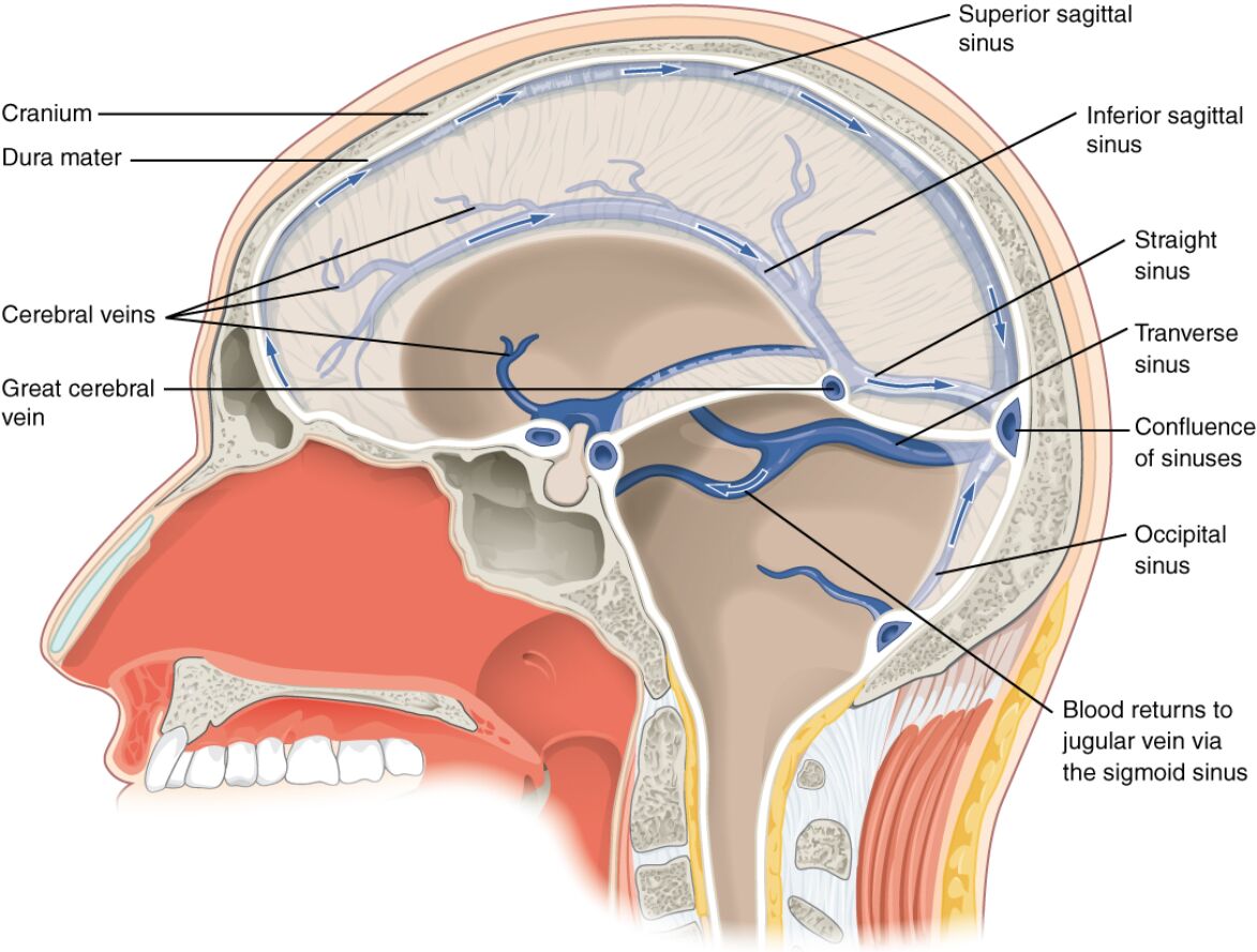
This section outlines each labeled component in the provided medical image, offering a detailed explanation of their anatomical roles and significance.
Cranium The cranium forms the bony structure encasing the brain, providing protection and support for the dural sinuses and cerebral veins. It consists of several fused bones, such as the frontal, parietal, and occipital bones, which create a stable environment for intracranial structures.
Dura mater The dura mater is the outermost layer of the meninges, a tough fibrous membrane that envelops the brain and spinal cord, housing the dural sinuses. It provides structural integrity and serves as a conduit for venous drainage, with its inner layer reflecting to form sinus walls.
Cerebral veins The cerebral veins collect blood from the brain parenchyma, draining into the dural sinuses to facilitate the removal of carbon dioxide and metabolic byproducts. These veins lack muscular walls, relying on the pressure gradient created by arterial inflow to propel blood toward the sinuses.
Great cerebral vein The great cerebral vein, also known as the vein of Galen, converges deep within the brain, collecting blood from the internal cerebral veins and choroid plexuses. It joins the inferior sagittal sinus to form the straight sinus, playing a key role in deep venous drainage.
Superior sagittal sinus The superior sagittal sinus runs along the midline of the cranial vault within the dura mater, receiving blood from the superior cerebral veins and arachnoid granulations. It drains into the confluence of sinuses, serving as a major pathway for venous return from the upper brain regions.
Inferior sagittal sinus The inferior sagittal sinus travels along the inferior edge of the falx cerebri, collecting blood from the falx and medial cerebral surfaces. It merges with the great cerebral vein to form the straight sinus, contributing to the posterior venous drainage system.
Straight sinus The straight sinus is formed by the union of the great cerebral vein and inferior sagittal sinus, running along the tentorium cerebelli. It drains blood from the cerebellum and deep brain structures, eventually joining the confluence of sinuses.
Transverse sinus The transverse sinus extends laterally from the confluence of sinuses along the occipital bone, receiving blood from the superior sagittal and straight sinuses. It continues as the sigmoid sinus, facilitating the transition of venous blood toward the jugular veins.
Confluence of sinuses The confluence of sinuses, located near the occipital bone, is the junction where the superior sagittal, straight, and occipital sinuses converge. This critical point directs blood flow into the transverse sinuses, ensuring efficient venous drainage from multiple regions.
Occipital sinus The occipital sinus runs along the midline of the occipital bone within the falx cerebelli, draining blood from the posterior fossa and cerebellum. It connects with the confluence of sinuses, providing an additional pathway for venous return in the posterior cranial region.
Blood returns to jugular vein via the sigmoid sinus The blood returns to jugular vein via the sigmoid sinus represents the final stage of cerebral venous drainage, where the transverse sinus narrows into the sigmoid sinus. This S-shaped channel directs deoxygenated blood into the internal jugular vein, completing the circulatory loop.
Anatomy of Dural Sinuses and Cerebral Veins
The dural sinuses and cerebral veins form a complex network that ensures the efficient removal of blood from the brain. This system is integral to maintaining intracranial homeostasis and supporting cerebral metabolism.
- Anatomically, the dural sinuses are endothelial-lined spaces between the periosteal and meningeal layers of the dura mater, devoid of valves to allow bidirectional flow.
- The cerebral veins, including superficial and deep systems, drain into these sinuses, with the superficial veins following the cortical surface and the deep veins converging toward the great cerebral vein.
- The dura mater’s rigidity helps contain the sinuses, preventing collapse under varying intracranial pressures.
- Variations in sinus anatomy, such as hypoplasia of the transverse sinus, are common and can influence venous drainage patterns.
Physiological Role in Venous Drainage
The dural sinuses and cerebral veins are essential for returning deoxygenated blood to the systemic circulation while managing intracranial pressure. Their function is closely tied to cerebral blood flow dynamics and pressure regulation.
- Physiologically, these structures handle approximately 600-700 ml of blood per minute, with the superior sagittal sinus accounting for a significant portion of this volume.
- The arachnoid granulations along the superior sagittal sinus filter cerebrospinal fluid (CSF) into the venous system, aiding in its absorption and pressure balance.
- Venous outflow is passive, driven by the pressure gradient between the brain and the right atrium, modulated by posture and respiratory cycles.
- In cases of venous sinus thrombosis, impaired drainage can lead to increased intracranial pressure, necessitating prompt intervention.
Clinical Relevance and Imaging
Understanding the anatomy and function of dural sinuses and cerebral veins is crucial for diagnosing and managing venous-related pathologies. Imaging techniques provide valuable insights into their structure and potential abnormalities.
- Clinically, conditions like cerebral venous sinus thrombosis can obstruct these sinuses, leading to symptoms such as headaches and seizures due to elevated intracranial pressure.
- Magnetic resonance venography (MRV) and computed tomography venography (CTV) are preferred imaging modalities to visualize sinus patency and detect thrombi.
- Anatomical variants, such as a dominant transverse sinus, may predispose individuals to uneven drainage, influencing surgical approaches.
- Preventive strategies include managing risk factors like dehydration and hypercoagulable states, which can precipitate venous thrombosis.
Blood Flow Pathways and Connections
The pathway of blood from the brain to the jugular veins involves a series of interconnected structures that ensure efficient drainage. This process highlights the integration of venous and dural systems in cerebral circulation.
- Blood from the cerebral cortex drains into the superficial cerebral veins, which empty into the superior sagittal sinus.
- Deep structures, including the basal ganglia and thalamus, are drained by the internal cerebral veins into the great cerebral vein.
- The confluence of sinuses acts as a central hub, distributing flow into the transverse and occipital sinuses based on pressure gradients.
- Ultimately, the sigmoid sinus channels blood into the internal jugular vein, where it returns to the heart via the superior vena cava.
In conclusion, the dural sinuses and cerebral veins represent a finely tuned system that supports the brain’s metabolic demands while safeguarding against pressure imbalances. Their anatomical complexity and physiological importance underscore the need for detailed study and clinical awareness, paving the way for advancements in neurovascular care.

