The Circle of Willis represents a critical arterial anastomosis at the base of the brain, formed by the interconnection of major arteries that supply oxygenated blood to neural tissues. This polygonal structure ensures collateral circulation, protecting the brain from ischemia during vascular occlusions or variations in blood flow. Comprising branches from the internal carotid arteries and vertebral arteries, it plays a pivotal role in maintaining cerebral perfusion, highlighting its significance in neurovascular anatomy and potential implications in conditions like strokes.
Labeled Structures in the Circle of Willis
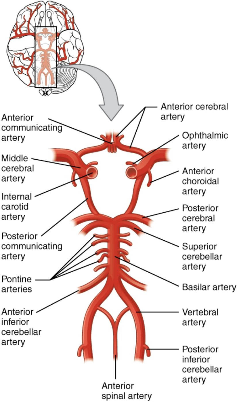
This section details each labeled artery in the provided medical image, offering insights into their origins, paths, and functional roles.
Anterior communicating artery The anterior communicating artery serves as a short connecting vessel between the left and right anterior cerebral arteries, facilitating blood flow equalization across hemispheres. It is typically located anteriorly in the Circle of Willis and can vary in size, sometimes presenting as a single trunk or duplicated segments.
Anterior cerebral artery The anterior cerebral artery arises from the internal carotid artery and courses medially to supply the medial and superior surfaces of the frontal and parietal lobes. It provides essential branches like the pericallosal and callosomarginal arteries, which nourish areas involved in motor control of the lower limbs and cognitive functions.
Ophthalmic artery The ophthalmic artery branches from the internal carotid artery just after it enters the cranial cavity, primarily supplying the orbit, eye structures, and parts of the nasal cavity. It plays a crucial role in visual health by perfusing the retina, optic nerve, and extraocular muscles through its central retinal and ciliary branches.
Middle cerebral artery The middle cerebral artery is the largest branch of the internal carotid artery, extending laterally to irrigate the lateral surfaces of the cerebral hemispheres, including key areas for language and sensory processing. Its lenticulostriate branches penetrate deeper to supply the basal ganglia and internal capsule, making it vital for motor and sensory pathways.
Internal carotid artery The internal carotid artery ascends from the common carotid artery in the neck, entering the skull via the carotid canal to deliver blood to the anterior brain circulation. It bifurcates into the anterior and middle cerebral arteries after giving off the ophthalmic and other smaller branches, forming a foundational component of cerebral blood supply.
Anterior choroidal artery The anterior choroidal artery originates from the internal carotid artery distal to the posterior communicating artery, supplying structures like the choroid plexus, optic tract, and parts of the hippocampus. Its perfusion territory includes critical regions such as the posterior limb of the internal capsule and the lateral geniculate body, influencing vision and memory functions.
Posterior cerebral artery The posterior cerebral artery emerges from the basilar artery, encircling the midbrain to supply the occipital lobe, inferior temporal lobe, and thalamus. It provides branches like the thalamoperforating and cortical arteries, essential for visual processing and sensory integration.
Posterior communicating artery The posterior communicating artery links the internal carotid artery to the posterior cerebral artery, acting as a bridge between anterior and posterior circulations. This vessel allows for compensatory blood flow in cases of arterial stenosis, though it can also be a site for aneurysms.
Superior cerebellar artery The superior cerebellar artery branches from the basilar artery near its termination, supplying the superior surface of the cerebellum, midbrain, and pineal gland. It contributes to coordination and balance by perfusing cerebellar cortex and deep nuclei, with potential impacts on ataxia if occluded.
Pontine arteries The pontine arteries consist of multiple small perforating branches from the basilar artery, directly supplying the pons and its neural tracts. These vessels ensure the integrity of brainstem functions, including cranial nerve nuclei and corticospinal pathways, critical for basic life-sustaining activities.
Basilar artery The basilar artery forms from the convergence of the two vertebral arteries at the base of the pons, ascending along the ventral brainstem to bifurcate into the posterior cerebral arteries. It distributes blood to the brainstem, cerebellum, and posterior brain via its paramedian and circumferential branches.
Anterior inferior cerebellar artery The anterior inferior cerebellar artery typically arises from the basilar artery, supplying the anterior and inferior portions of the cerebellum, as well as parts of the pons and inner ear structures. It supports functions like equilibrium and hearing through its labyrinthine branch, which vascularizes the cochlea and vestibular apparatus.
Vertebral artery The vertebral artery originates from the subclavian artery, ascending through the cervical vertebrae to enter the skull and merge into the basilar artery. It provides posterior circulation to the medulla, cerebellum, and spinal cord, with segments that can be prone to dissection in trauma.
Posterior inferior cerebellar artery The posterior inferior cerebellar artery branches from the vertebral artery, irrigating the inferior cerebellar surface, choroid plexus of the fourth ventricle, and lateral medulla. It is involved in maintaining postural stability and can be associated with Wallenberg syndrome if infarcted.
Anterior spinal artery The anterior spinal artery forms from branches of the vertebral arteries, descending along the anterior median fissure of the spinal cord to supply its anterior two-thirds. This artery ensures motor neuron viability and sensory tract function, with occlusions potentially leading to anterior spinal artery syndrome characterized by paraplegia and loss of pain sensation.
Anatomy and Formation of the Circle of Willis
The Circle of Willis is an arterial ring that encircles the optic chiasm and pituitary stalk, providing a redundant blood supply system to mitigate risks from vascular disruptions. Its formation involves contributions from both carotid and vertebrobasilar systems, creating a hexagonal or polygonal configuration in most individuals.
- Embryologically, the Circle of Willis develops from the fusion of primitive arterial networks during fetal growth, with variations occurring in up to 50% of the population, such as hypoplasia of certain segments.
- Anatomically, it integrates the anterior circulation from the internal carotid arteries with the posterior circulation from the vertebral-basilar system, ensuring balanced hemodynamics.
- Key junctions include the anterior and posterior communicating arteries, which allow for cross-flow during unilateral obstructions.
- Imaging techniques like magnetic resonance angiography (MRA) or computed tomography angiography (CTA) are commonly used to visualize its structure and detect anomalies.
Physiological Role in Cerebral Circulation
Blood flow through the Circle of Willis maintains constant cerebral perfusion pressure, adapting to changes in systemic blood pressure via autoregulation mechanisms. This collateral pathway is essential for neuroprotection, as it can reroute blood in response to occlusions, preserving brain function.
- Physiologically, the arteries deliver oxygen and nutrients while removing metabolic waste, with flow rates averaging 700-800 ml/min in adults.
- The internal carotid arteries contribute approximately 80% of cerebral blood, while the vertebral arteries provide the remaining 20%, converging at the basilar artery.
- Autoregulation involves myogenic responses in arterial smooth muscle, maintaining flow between 50-150 mmHg mean arterial pressure.
- In conditions of hypotension, the circle’s anastomoses become crucial, preventing hypoperfusion in vulnerable watershed areas.
Clinical Significance and Variations
Variations in the Circle of Willis anatomy can influence susceptibility to cerebrovascular events, with incomplete circles increasing risks in certain populations. Understanding these differences aids in preoperative planning for neurosurgical interventions and stroke management strategies.
- Common variants include aplasia of the posterior communicating artery or fetal-type posterior cerebral artery, where it arises directly from the internal carotid.
- In clinical practice, aneurysms often form at bifurcation points within the circle, such as the anterior communicating artery, due to hemodynamic stress.
- Diagnostic tools like Doppler ultrasound assess flow dynamics, while endovascular treatments target pathologies within these vessels.
- Preventive measures focus on controlling risk factors like hypertension and atherosclerosis, which can compromise the circle’s integrity.
Blood Supply Pathways to the Brain
The brain’s blood supply enters via the internal carotid and vertebral arteries, forming intricate networks that ensure comprehensive coverage of neural regions. These pathways are designed for efficiency, with branching patterns optimized for minimal resistance and maximal distribution.
- From the neck, the internal carotid arteries travel through the petrous bone, cavernous sinus, and sphenoid bone before entering the subarachnoid space.
- The vertebral arteries pierce the dura at the foramen magnum, uniting to form the basilar artery on the clivus.
- Terminal branches like the anterior, middle, and posterior cerebral arteries divide the cerebrum into vascular territories, each with specific functional associations.
- Smaller perforators, such as lenticulostriate and thalamogeniculate arteries, supply deep structures like the basal ganglia and thalamus.
In summary, the Circle of Willis exemplifies the body’s ingenious design for safeguarding cerebral function through redundant vascular pathways. Its detailed anatomy and physiology underscore the importance of vascular health in preventing neurological deficits, encouraging ongoing research into neurovascular dynamics.

