The action potential in cardiac contractile cells is a critical process that drives the heart’s rhythmic contractions, distinctly different from skeletal muscle due to its unique phases. This chart illustrates the long plateau phase and extended refractory period caused by calcium ion influx, while comparing it to skeletal muscle action potential, offering a clear view of cardiac electrophysiology. Exploring this image provides valuable insights into how these cells sustain the heart’s pumping action.
Labelled Parts Explanation
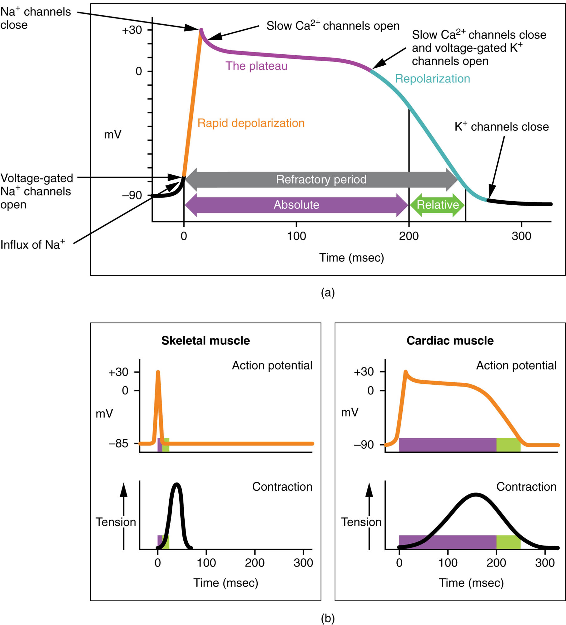
- Long plateau phase The long plateau phase is a prolonged depolarization period due to a significant influx of calcium ions through L-type calcium channels. It maintains the contractile cells’ membrane potential, allowing sustained contraction of the heart muscle.
- Extended refractory period The extended refractory period follows the plateau phase, preventing another action potential until the cell fully relaxes, due to a long absolute refractory period. This ensures the heart completes its contraction and refilling before the next beat, avoiding tetanus.
- Cardiac muscle action potential The cardiac muscle action potential depicts the electrical changes in contractile cells, featuring rapid depolarization, a prolonged plateau, and repolarization phases. It differs from skeletal muscle by its dependence on calcium for the plateau and its longer duration.
- Skeletal muscle action potential The skeletal muscle action potential shows a brief depolarization and repolarization without a plateau, relying solely on sodium and potassium ion movements. It is shorter in duration and lacks the refractory period extension seen in cardiac cells.
Anatomical Overview of Cardiac Contractile Cell Action Potential
The action potential in cardiac contractile cells is tailored for the heart’s continuous pumping needs. This chart highlights the phases that distinguish cardiac from skeletal muscle activity.
- The long plateau phase sustains contraction, driven by calcium influx.
- The extended refractory period prevents re-excitation, ensuring complete relaxation.
- The cardiac muscle action potential reflects the heart’s rhythmic demands.
- The skeletal muscle action potential serves as a contrast, showing a simpler electrical profile.
This comparison underscores the heart’s specialized electrophysiology.
Role of the Long Plateau Phase
The long plateau phase is a defining feature of cardiac action potentials. Its duration supports effective contraction.
- The long plateau phase results from calcium entering through voltage-gated channels.
- This influx triggers the release of additional calcium from the sarcoplasmic reticulum.
- The prolonged depolarization maintains muscle tension during systole.
- This phase distinguishes cardiac cells from the brief skeletal muscle action.
The plateau ensures sustained pumping action.
Significance of the Extended Refractory Period
The extended refractory period prevents overstimulation of the heart. This feature protects cardiac function.
- The extended refractory period lasts nearly as long as the contraction, due to slow calcium channel closure.
- It includes a long absolute refractory period, blocking premature beats.
- This prevents tetanic contraction, which would impair blood flow.
- The period aligns with the mechanical cycle for efficient pumping.
This mechanism safeguards the heart’s rhythm.
Comparison of Cardiac and Skeletal Muscle Action Potentials
The cardiac and skeletal muscle action potentials differ in structure and function. This chart highlights their unique profiles.
- The cardiac muscle action potential features a plateau due to calcium influx, lasting 200-300 ms.
- The skeletal muscle action potential is brief, around 2-5 ms, relying on sodium influx alone.
- The long plateau phase in cardiac cells supports sustained contraction.
- The extended refractory period is absent in skeletal muscle, allowing rapid successive contractions.
This contrast reflects their distinct physiological roles.
Ionic Basis of Cardiac Action Potential
The action potential in cardiac contractile cells depends on specific ion movements. These shifts drive the electrical and mechanical events.
- The long plateau phase is sustained by calcium influx through L-type channels.
- Rapid depolarization begins with sodium influx, similar to skeletal muscle.
- The extended refractory period involves slow potassium efflux during repolarization.
- Calcium release from the sarcoplasmic reticulum amplifies contraction strength.
This ionic dance is key to cardiac performance.
Physiological Importance of the Action Potential
The action potential’s design optimizes the heart’s pumping efficiency. Its phases support continuous circulation.
- The long plateau phase ensures adequate time for ventricular ejection.
- The extended refractory period prevents arrhythmias by blocking re-excitation.
- The cardiac muscle action potential’s duration matches the mechanical cycle.
- This synchronization maintains steady blood flow.
The process is vital for cardiovascular stability.
Clinical Relevance of Cardiac Action Potential
Understanding the cardiac action potential aids in diagnosing heart conditions. These phases are key clinical indicators.
- Prolongation of the long plateau phase can lead to long QT syndrome, risking torsades de pointes.
- Shortening the extended refractory period may cause re-entrant arrhythmias.
- The cardiac muscle action potential’s profile is assessed via electrocardiograms.
- Treatments target ion channel function to normalize these phases.
This knowledge guides effective cardiac management.
Conclusion
The action potential in cardiac contractile cells chart provides a detailed exploration of the electrical events that drive the heart’s rhythmic contractions. By examining the long plateau phase, extended refractory period, cardiac muscle action potential, and its comparison to the skeletal muscle action potential, one gains insight into the heart’s unique physiology. This understanding serves as a foundation for studying cardiovascular function and addressing related health issues, encouraging further exploration of the heart’s intricate electrical and mechanical design.

