The human circulatory system operates through a dual mechanism, comprising the pulmonary and systemic circuits, to ensure efficient oxygen and nutrient delivery. This article explores the pathways illustrated in the diagram, tracing blood flow from the right atrium through the pulmonary circuit for oxygenation, then to the left ventricle for systemic distribution. Delving into this process reveals the heart’s coordinated role in maintaining bodily homeostasis.
Right atrium
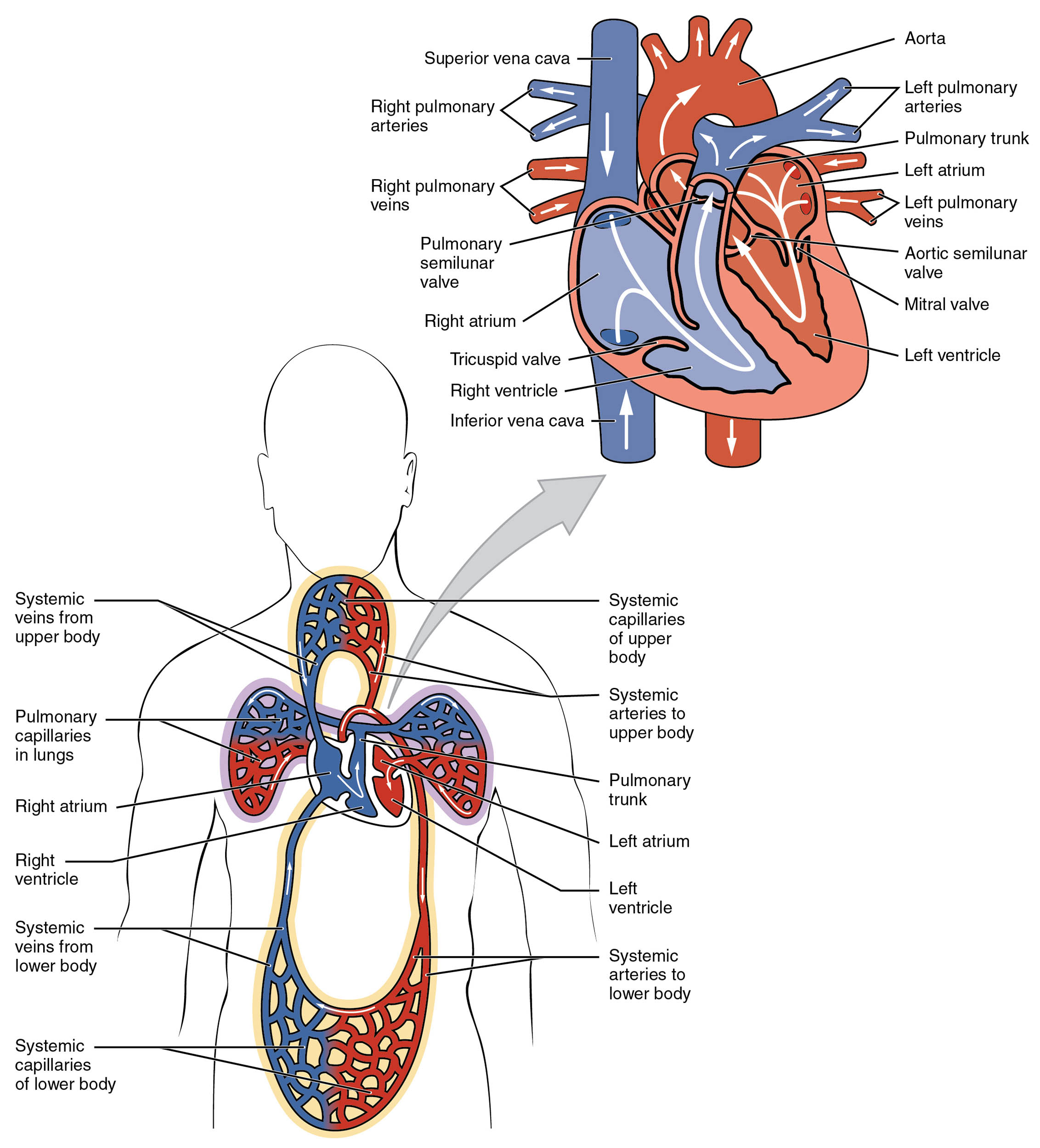
- The right atrium receives deoxygenated blood from the body via the superior and inferior vena cava.
- It pumps this blood into the right ventricle to begin the pulmonary circulation.
Right ventricle
- The right ventricle receives blood from the right atrium and pumps it into the pulmonary artery.
- Its thinner walls are adapted to handle the lower pressure of pulmonary circulation.
Pulmonary artery
- The pulmonary artery carries deoxygenated blood from the right ventricle to the lungs for gas exchange.
- It branches into smaller vessels, delivering blood to the pulmonary capillaries.
Pulmonary capillaries
- Pulmonary capillaries are tiny vessels in the lungs where gas exchange occurs, with oxygen entering the blood and carbon dioxide being removed.
- Their thin walls facilitate the diffusion of gases between the alveoli and bloodstream.
Pulmonary veins
- Pulmonary veins return oxygenated blood from the lungs to the left atrium.
- They are unique among veins as they carry oxygen-rich blood, unlike most veins.
Left atrium
- The left atrium receives oxygenated blood from the pulmonary veins and prepares it for systemic circulation.
- It contracts to push blood into the left ventricle.
Left ventricle
- The left ventricle pumps oxygenated blood into the aorta for distribution through the systemic circuit.
- Its thick, muscular walls generate the high pressure needed for systemic circulation.
Aorta
- The aorta is the largest artery, carrying oxygenated blood from the left ventricle to the rest of the body.
- It branches into major arteries, ensuring blood reaches all tissues.
Systemic capillaries
- Systemic capillaries facilitate the exchange of oxygen, nutrients, and waste products between blood and body tissues.
- Their extensive network allows for efficient delivery and removal of substances.
Systemic veins
- Systemic veins collect deoxygenated blood from the body’s tissues and return it to the right atrium.
- They include the superior and inferior vena cava as major return pathways.
The dual system of human blood circulation is a remarkable process that sustains life by oxygenating blood and delivering it to tissues. The diagram illustrates the journey from the right atrium, where deoxygenated blood enters, through the pulmonary artery to the lungs for oxygenation in the pulmonary capillaries, and back via the pulmonary veins to the left atrium. From there, the left ventricle propels the oxygenated blood into the aorta for systemic distribution, with systemic capillaries facilitating exchange before systemic veins return it to the right atrium, restarting the cycle.
Pulmonary Circulation: Oxygenating the Blood
Pulmonary circulation begins in the right side of the heart, focusing on oxygenating deoxygenated blood. This circuit is essential for preparing blood for systemic use.
- The right atrium and right ventricle work together to send blood into the pulmonary artery.
- In the pulmonary capillaries, oxygen from the alveoli diffuses into the blood, while carbon dioxide is expelled.
Systemic Circulation: Delivering Oxygen to Tissues
Systemic circulation distributes oxygenated blood from the left side of the heart to the body’s tissues. This process ensures vital nutrients and oxygen reach every cell.
- The left atrium receives oxygenated blood and passes it to the left ventricle for pumping into the aorta.
- Systemic capillaries then release oxygen and nutrients while collecting carbon dioxide and wastes for return.
The human circulatory system is ingeniously divided into pulmonary and systemic circuits, each serving distinct yet interconnected roles. The pulmonary circulation starts as deoxygenated blood enters the right atrium from the superior and inferior vena cava, reflecting the body’s venous return. The right ventricle, with its thinner muscular walls suited for lower pressure, pumps this blood into the pulmonary artery, which carries it to the lungs. Within the pulmonary capillaries, gas exchange occurs: oxygen diffuses from the alveoli into the blood, raising its oxygen saturation, while carbon dioxide, a metabolic byproduct, is released into the alveoli for exhalation. The pulmonary veins then transport this newly oxygenated blood back to the left atrium, completing the pulmonary loop.
The systemic circulation begins as the left atrium contracts, pushing blood into the left ventricle, which has thick, muscular walls to generate the high pressure needed for arterial distribution. The aorta, the body’s largest artery, receives this blood and branches into systemic arteries, delivering oxygen and nutrients to tissues via the systemic capillaries. In these capillaries, oxygen and glucose exit to nourish cells, while carbon dioxide and waste products like urea enter the bloodstream. The systemic veins, including the superior and inferior vena cava, collect this deoxygenated blood and return it to the right atrium, perpetuating the cycle. This dual system ensures a continuous supply of oxygenated blood and efficient waste removal.
The heart’s structure supports this dual circulation, with the right side handling pulmonary flow and the left side managing systemic pressure. The pulmonary artery is the only artery carrying deoxygenated blood, a unique adaptation for lung-bound travel, while the pulmonary veins are exceptional veins carrying oxygenated blood. The left ventricle’s robust musculature, approximately three times thicker than the right ventricle’s, reflects the higher pressure required to pump blood through the systemic circuit, which spans the entire body. Cardiac output, typically 5 liters per minute at rest, adjusts dynamically to meet metabolic demands, regulated by the autonomic nervous system.
The capillary networks—pulmonary capillaries and systemic capillaries—are critical for exchange efficiency. Pulmonary capillaries, with a surface area of about 70-100 square meters, maximize gas exchange due to their proximity to alveolar air sacs. Systemic capillaries, numbering around 10 billion, ensure nutrient delivery and waste removal across all tissues, from the brain to the extremities. The pressure gradient drives this flow, with pulmonary circulation operating at 15-25 mmHg and systemic circulation at 120/80 mmHg, highlighting the heart’s dual-pressure system. Valves within the heart, such as the tricuspid and mitral valves, prevent backflow, maintaining directional flow.
This circulatory design supports homeostasis by balancing oxygen supply and carbon dioxide removal. The aorta’s elastic walls smooth out the pulsatile flow from the left ventricle, ensuring steady delivery, while the systemic veins’ larger lumens accommodate the returning blood volume. The system’s efficiency can be assessed via metrics like cardiac index or pulmonary artery pressure, aiding in diagnosing conditions like pulmonary hypertension or heart failure. The interplay between these circuits underscores the heart’s role as a dual pump, adapting to exercise or rest through increased heart rate and stroke volume.
The dual circulatory system exemplifies the body’s intricate design for survival. By separating pulmonary and systemic flows, the heart ensures oxygenated and deoxygenated blood remain distinct, optimizing tissue perfusion. This understanding not only enhances appreciation for cardiovascular physiology but also informs clinical approaches to maintaining circulatory health.

