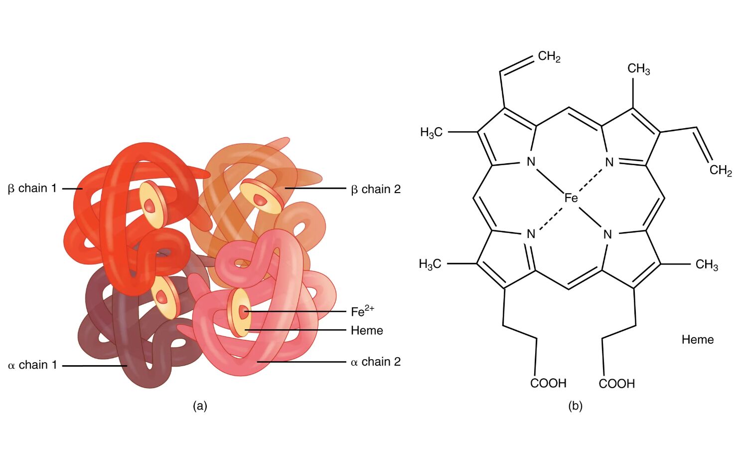
Hemoglobin is a vital protein in red blood cells, responsible for oxygen transport throughout the body and playing a key role in maintaining acid-base balance. This diagram illustrates the intricate structure of a hemoglobin molecule, highlighting its four globin protein chains and heme groups, which enable its oxygen-carrying capacity. Understanding this molecular architecture provides insight into its physiological significance and the processes it supports.
Key Components of the Hemoglobin Molecule
The diagram breaks down the hemoglobin structure into its essential parts.
α chain 1:
This is one of the two alpha globin chains that form part of the hemoglobin tetramer, contributing to the protein’s overall stability and oxygen-binding sites. Each alpha chain binds to a heme group, facilitating the molecule’s ability to transport oxygen efficiently.
α chain 2:
The second alpha globin chain mirrors α chain 1, forming a symmetrical pair that enhances hemoglobin’s quaternary structure. Together with the beta chains, it ensures the cooperative binding of oxygen molecules.
β chain 1:
This beta globin chain is one of two that pair with the alpha chains, playing a crucial role in oxygen affinity and allosteric regulation. It contains a heme-binding pocket, essential for the protein’s function in gas exchange.
β chain 2:
The second beta globin chain complements β chain 1, contributing to the hemoglobin molecule’s ability to adapt its oxygen-binding capacity based on physiological conditions. It interacts with alpha chains to form a functional tetramer.
Heme:
Heme is the iron-containing pigment bound to each globin chain, featuring a ferrous iron (Fe²⁺) ion that binds oxygen reversibly. This structure, depicted in detail, is critical for hemoglobin’s role in oxygen transport and carbon dioxide removal.
Fe²⁺:
The ferrous iron ion within the heme group is the site where oxygen molecules bind, undergoing a conformational change that enhances further oxygen uptake. Its presence in the reduced state is essential for effective gas transport.
The Anatomical and Physiological Role of Hemoglobin
Hemoglobin’s structure is a marvel of biological engineering, enabling red blood cells to carry oxygen from the lungs to tissues and return carbon dioxide for exhalation. The tetrameric arrangement of two alpha and two beta chains, each associated with a heme group, allows for cooperative binding, where the binding of one oxygen molecule increases the affinity for additional molecules. This allosteric effect, influenced by pH and carbon dioxide levels via the Bohr effect, ensures efficient oxygen delivery in varying physiological states.
The heme group’s Fe²⁺ ion forms a reversible bond with oxygen, a process facilitated by the globin chains’ hydrophobic pockets. Hemoglobin also transports a small amount of carbon dioxide as carbaminohemoglobin, aiding in acid-base balance alongside the chloride shift. Hormones like erythropoietin, released by the kidneys in response to hypoxia, stimulate red blood cell production to maintain adequate hemoglobin levels.
- Oxygen Transport: Each hemoglobin molecule can bind up to four oxygen molecules; affinity decreases in acidic environments like active muscles.
- Carbon Dioxide Role: About 10% of CO₂ is carried as carbaminohemoglobin; the rest is transported as bicarbonate ions.
- Structural Stability: Alpha and beta chains are encoded by different genes, with mutations leading to variants like hemoglobin S in sickle cell disease.
Physical Characteristics and Clinical Relevance
The physical structure of hemoglobin, as shown, reflects its functional efficiency, with a compact globular shape that fits within erythrocytes. A single red blood cell contains approximately 300 million hemoglobin molecules, enabling the transport of over 1 billion oxygen molecules, a testament to its high capacity.
Clinically, hemoglobin levels are measured to assess oxygen-carrying capacity, with abnormalities indicating conditions like anemia or polycythemia. The diagram’s depiction of heme and Fe²⁺ is crucial for understanding disorders such as thalassemia, where defective globin chain synthesis reduces hemoglobin function. Electrophoresis can identify hemoglobin variants, guiding treatments like blood transfusions or iron supplementation.
- Diagnostic Tools: Hemoglobin A1c tests reflect average blood glucose over months; pulse oximetry monitors oxygen saturation.
- Therapeutic Approaches: Folic acid and vitamin B12 correct megaloblastic anemia; hydroxyurea increases fetal hemoglobin in sickle cell disease.
Conclusion
The hemoglobin molecule diagram reveals the elegant design that underpins oxygen delivery and carbon dioxide removal, showcasing the interplay of globin chains and heme groups. This structure not only supports vital physiological processes but also serves as a diagnostic marker for various hematological conditions. By exploring its components, one gains a deeper appreciation for the body’s ability to adapt and thrive, emphasizing the importance of maintaining optimal hemoglobin function.

