The cerebral cortex is a vital component of the human brain, renowned for its extensive folding that maximizes surface area for complex functions. This image of the lobes of the cerebral cortex highlights key regions, including the frontal lobe, precentral gyrus, central sulcus, parietal lobe, postcentral gyrus, parieto-occipital sulcus, occipital lobe, temporal lobe, and lateral sulcus, offering a detailed view of its anatomical layout. This article explores the structure and function of these lobes, providing an in-depth understanding of their roles in the central nervous system.
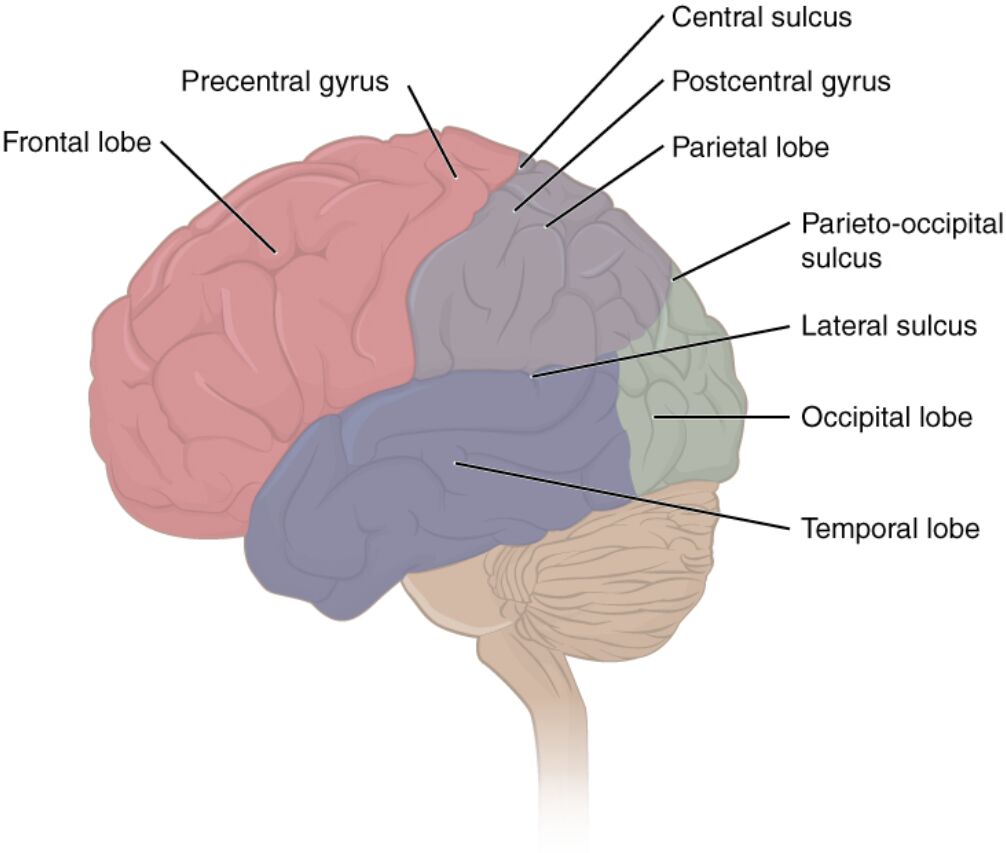
Frontal lobe
The frontal lobe, located at the front of the brain, is responsible for higher cognitive functions such as decision-making, problem-solving, and planning. It also houses the primary motor cortex, which controls voluntary movements.
Precentral gyrus
The precentral gyrus lies within the frontal lobe and serves as the primary motor cortex, orchestrating precise voluntary movements. Damage to this area can result in motor deficits, affecting coordinated actions.
Central sulcus
The central sulcus is a prominent groove that separates the frontal lobe from the parietal lobe, marking the boundary between motor and sensory regions. This sulcus plays a critical role in organizing the brain’s functional map.
Parietal lobe
The parietal lobe, situated behind the frontal lobe, processes sensory information such as touch, temperature, and pain. It also contributes to spatial orientation and integration of sensory data.
Postcentral gyrus
The postcentral gyrus, found in the parietal lobe, functions as the primary somatosensory cortex, receiving and interpreting tactile sensations. It maps the body’s sensory input, allowing for spatial awareness.
Parieto-occipital sulcus
The parieto-occipital sulcus delineates the boundary between the parietal and occipital lobes, aiding in the structural organization of the cortex. This sulcus helps define the visual processing regions of the brain.
Occipital lobe
The occipital lobe, located at the back of the brain, is primarily responsible for visual processing and interpretation. It contains the primary visual cortex, which receives and analyzes visual information from the eyes.
Temporal lobe
The temporal lobe, positioned on the sides of the brain, is involved in auditory processing, memory, and language comprehension. It houses the primary auditory cortex and structures like the hippocampus for memory formation.
Lateral sulcus
The lateral sulcus, also known as the Sylvian fissure, separates the temporal lobe from the frontal and parietal lobes. This deep groove accommodates important neural pathways and contributes to brain stability.
Anatomical Overview of the Cerebral Cortex
The cerebral cortex is the brain’s outer layer, characterized by its folded structure that enhances its functional capacity. This folding, visible in the lobes, supports a vast network of neural activity.
- The cortex is approximately 2-4 mm thick and contains six layers of neurons, varying in density and function.
- Its division into four main lobes—frontal, parietal, occipital, and temporal—allows for specialized processing of sensory, motor, and cognitive tasks.
- The sulci and gyri, such as the central and lateral sulcus, increase surface area, housing up to 16 billion neurons.
- Blood supply, primarily from the middle cerebral artery, ensures the cortex receives adequate oxygen and glucose for its high metabolic demand.
Detailed Exploration of the Frontal Lobe and Precentral Gyrus
The frontal lobe and precentral gyrus are central to executive function and movement. Their proximity enhances their coordinated role in behavior and action.
- The frontal lobe includes the prefrontal cortex, which regulates personality, social behavior, and complex planning.
- The precentral gyrus contains a somatotopic map, where different body parts are represented in specific cortical areas.
- Damage to the frontal lobe can lead to changes in personality or impaired judgment, as seen in frontal lobe syndrome.
- The precentral gyrus receives input from the basal ganglia and cerebellum, refining motor control.
Understanding the Parietal and Occipital Lobes
The parietal and occipital lobes handle sensory integration and visual processing, respectively, forming a critical sensory hub. Their distinct roles support perception and spatial awareness.
- The parietal lobe integrates sensory data from the postcentral gyrus, contributing to body schema and spatial navigation.
- The occipital lobe’s primary visual cortex processes raw visual data, relayed via the optic nerve and lateral geniculate nucleus.
- The parieto-occipital sulcus serves as a landmark for neurosurgical procedures targeting these regions.
- Lesions in the occipital lobe can cause visual field defects, such as homonymous hemianopia.
Insights into the Temporal Lobe and Sulci
The temporal lobe and associated sulci play a key role in auditory and memory functions. Their structure supports complex cognitive processes.
- The temporal lobe contains the hippocampus, essential for forming new memories and spatial memory.
- The lateral sulcus houses the insular cortex, involved in taste, interoception, and autonomic functions.
- The central sulcus separates motor and sensory areas, influencing the temporal lobe’s auditory processing.
- Temporal lobe epilepsy can arise from abnormal electrical activity, often managed with medication or surgery.
Clinical Relevance and Functional Importance
The lobes of the cerebral cortex are critical for diagnosing and treating neurological conditions. Their specialized functions guide clinical interventions.
- Damage to the frontal lobe may result in motor aphasia if Broca’s area is affected, impacting speech production.
- The parietal lobe’s role in spatial awareness is assessed in neglect syndrome, common after stroke.
- Occipital lobe injuries can lead to cortical blindness, requiring visual rehabilitation strategies.
- Imaging modalities like fMRI map cortical activity, aiding in epilepsy surgery planning.
Conclusion
The lobes of the cerebral cortex, as depicted in this image, reveal the brain’s remarkable specialization and interconnectedness. From the frontal lobe’s role in decision-making to the occipital lobe’s visual processing, each region contributes uniquely to human cognition and sensory experience. Exploring their anatomy not only deepens our understanding of brain function but also enhances approaches to neurological care and research.

