nervous system, somatic nervous system, autonomic nervous system, enteric nervous system, central nervous system, peripheral nervous system, brain function, spinal cord, sensory neurons, motor neurons, ganglia, digestive tract, homeostasis, reflexes, voluntary movement, involuntary control, cranial nerves, spinal nerves, sympathetic ganglia, parasympathetic ganglia
The human nervous system is a complex network that coordinates actions and sensory information by transmitting signals to and from different parts of the body. Divided into the central nervous system (CNS) and peripheral nervous system (PNS), it encompasses somatic, autonomic, and enteric structures that work together to maintain bodily functions, from conscious movements to unconscious regulation of internal organs.
Key Labeled Components in the Nervous System Diagram
This section breaks down each labeled part of the image, providing clear explanations of their roles in somatic, autonomic, and enteric functions.
Brain (CNS)
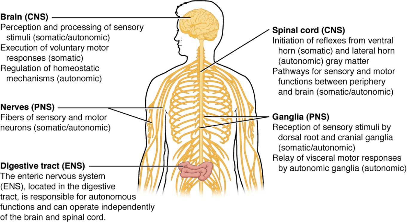
The brain, as part of the CNS, is responsible for the perception and processing of sensory stimuli, handling both somatic and autonomic inputs to interpret environmental and internal signals. It executes voluntary motor responses through somatic pathways and regulates homeostatic mechanisms via autonomic controls, ensuring balanced physiological states like temperature and heart rate.
Spinal Cord (CNS)
The spinal cord initiates reflexes from the ventral horn for somatic actions and the lateral horn for autonomic responses, allowing quick reactions without brain involvement. It serves as a conduit for sensory and motor pathways between the periphery and the brain, facilitating both somatic and autonomic functions essential for coordinated body movements and organ regulation.
Nerves (PNS)
Nerves in the PNS consist of fibers from sensory and motor neurons that transmit somatic and autonomic signals, connecting the CNS to muscles, glands, and sensory receptors. These structures enable the relay of information for voluntary actions like walking and involuntary processes such as digestion, ensuring seamless communication across the body.
Ganglia (PNS)
Ganglia receive sensory stimuli through dorsal root and cranial ganglia for somatic and autonomic sensations, acting as relay points for peripheral signals to the CNS. They also facilitate the relay of visceral motor responses via autonomic ganglia, which modulate organ functions independently of conscious control.
Digestive Tract (ENS)
The enteric nervous system within the digestive tract manages autonomous functions like peristalsis and secretion, operating independently of the brain and spinal cord for efficient gastrointestinal activity. It integrates sensory inputs from the gut and coordinates motor outputs to maintain digestive homeostasis, influenced but not directly controlled by autonomic inputs.
Anatomical Overview of the Nervous System Divisions
The nervous system is fundamentally divided into the CNS, comprising the brain and spinal cord, and the PNS, which includes all nerves and ganglia outside the CNS. This division allows for specialized control over bodily functions, with further subdivisions into somatic and autonomic components for precise regulation.
- The CNS acts as the command center, integrating sensory data and issuing motor commands.
- Within the CNS, the brain processes higher-order functions like cognition and emotion, while the spinal cord handles reflex arcs and basic signal transmission.
- The PNS extends the CNS’s reach, with sensory fibers carrying information inward and motor fibers sending commands outward.
- Somatic structures primarily deal with external interactions, such as touch and voluntary muscle control.
- Autonomic structures focus on internal viscera, regulating heart rate, blood pressure, and glandular secretions without conscious effort.
- The enteric division, often considered a “second brain,” specializes in gastrointestinal control, featuring its own neurons and circuits.
Physiological Roles of Somatic Structures
Somatic structures enable interaction with the external environment through conscious control and sensory feedback. They are crucial for movement, posture, and perception of stimuli like pain, temperature, and pressure.
- Spinal nerves carry both motor and sensory fibers, allowing for precise control of skeletal muscles.
- Sensory ganglia, including posterior root ganglia and cranial nerve ganglia, house cell bodies of sensory neurons that detect changes in the environment.
- Voluntary motor responses originate from the brain’s motor cortex, traveling down the spinal cord to activate muscles via alpha motor neurons.
- Reflexes, such as the knee-jerk response, involve monosynaptic circuits in the spinal cord for rapid protection.
- Somatic sensory pathways ascend through the dorsal columns or spinothalamic tracts to reach the cerebral cortex for conscious awareness.
- Integration with autonomic functions occurs in areas like the hypothalamus, where somatic sensations can influence involuntary responses.
In-Depth Look at Autonomic Structures
The autonomic nervous system operates involuntarily, maintaining homeostasis through sympathetic and parasympathetic divisions. It responds to internal needs, adjusting organ functions in response to stress or rest.
- Sympathetic ganglia, located in chains along the spinal cord, prepare the body for “fight or flight” by increasing heart rate and dilating pupils.
- Parasympathetic ganglia, often near target organs, promote “rest and digest” activities, such as stimulating saliva production and slowing the heartbeat.
- Autonomic motor pathways involve preganglionic and postganglionic neurons, with acetylcholine and norepinephrine as key neurotransmitters.
- Sensory inputs from viscera travel via autonomic afferents to inform the CNS of internal states like blood pressure or organ distension.
- Homeostatic regulation includes control over endocrine glands; for instance, autonomic signals to the thyroid gland influence the release of hormones like T3 and T4 for metabolism.
- Integration with the enteric system allows for modulated gut activity during stress, where sympathetic activation slows digestion.
The Enteric Nervous System: Anatomy and Function
Embedded within the walls of the gastrointestinal tract, the enteric nervous system functions semi-independently to control digestion. It contains millions of neurons organized into plexuses that coordinate local responses.
- The myenteric plexus regulates motility, controlling the contraction and relaxation of smooth muscles for peristalsis.
- The submucosal plexus manages secretions and blood flow, ensuring optimal absorption and enzyme release.
- Enteric neurons include sensory, motor, and interneurons, forming reflex circuits that operate without CNS input.
- Influences from the autonomic system, via vagus and pelvic nerves, fine-tune enteric functions but do not override its autonomy.
- Neurotransmitters like serotonin and dopamine in the ENS modulate gut-brain communication, affecting mood and appetite.
- Pathophysiological implications arise when enteric dysfunction occurs, though this image focuses on normal anatomy.
Interconnections Between Nervous System Divisions
All divisions of the nervous system interconnect to ensure holistic body function. For example, somatic sensations can trigger autonomic responses, like sweating from heat.
- Pathways from the brain descend through the spinal cord to influence both somatic and autonomic effectors.
- Cranial nerves, such as the vagus nerve, bridge somatic and autonomic functions, carrying sensory data from the throat and motor signals to the heart.
- Ganglia serve as hubs: somatic ganglia for unipolar sensory neurons, autonomic for multipolar relay neurons.
- The ENS receives modulatory inputs from sympathetic and parasympathetic fibers, integrating with higher centers for appetite control.
- Reflex integration occurs at multiple levels, from spinal monosynaptic reflexes to complex enteric circuits.
- Physiological balance is maintained through feedback loops, where sensory ganglia detect changes and motor responses adjust accordingly.
Clinical Relevance and Physiological Insights
Understanding these structures aids in comprehending how disruptions can lead to disorders, though the focus here remains on normal function. Physiological processes like hormone regulation exemplify the system’s complexity.
- Thyroid hormone release (T3 and T4) is influenced by autonomic inputs to the pituitary-hypothalamic axis, affecting basal metabolic rate.
- Reflex initiation in the spinal cord’s ventral horn protects against injury, while lateral horn activity regulates visceral tone.
- Sensory processing in the brain involves somatic maps in the somatosensory cortex and autonomic centers in the brainstem.
- Motor execution ranges from fine somatic control in handwriting to broad autonomic adjustments in blood flow.
- Homeostasis relies on autonomic feedback, with ganglia amplifying signals for efficient response.
- The ENS’s independence allows digestion to continue during sleep or unconscious states, highlighting evolutionary adaptations.
In summary, the somatic, autonomic, and enteric structures form an integrated nervous system that supports life through sensory perception, motor control, and internal regulation. This diagram illustrates their interconnected roles, emphasizing the body’s ability to function both consciously and autonomously for overall health and adaptation.

