The nervous system is a complex network that governs communication throughout the body, divided into the central nervous system (CNS) and the peripheral nervous system (PNS). This anatomical image highlights key structures such as the brain, spinal cord, ganglion, and nerve, offering a clear view of their roles in coordinating sensory and motor functions. Understanding the distinct yet interconnected components of the CNS and PNS provides valuable insights into how the body processes information and responds to its environment.
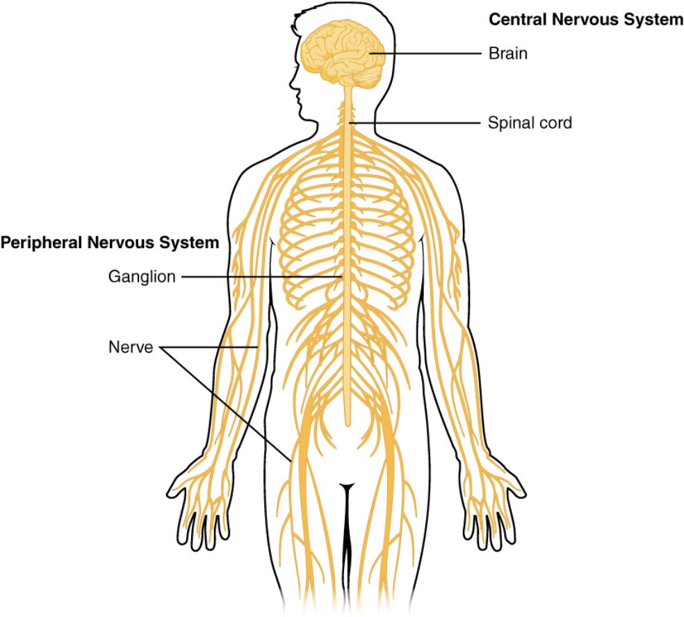
Labeled Components of the Nervous System
Brain: The brain serves as the primary control center of the central nervous system, housing regions like the cerebrum, cerebellum, and brainstem. It processes sensory input, initiates motor responses, and regulates vital functions such as breathing and heart rate.
Spinal cord: The spinal cord extends from the brainstem down the vertebral column, acting as a relay between the brain and the body. It facilitates reflex actions and transmits neural signals to and from the peripheral nerves.
Central Nervous System: This system encompasses the brain and spinal cord, forming the core of neural integration and command. Its structures are best studied microscopically in prepared tissue due to their intricate cellular organization.
Peripheral Nervous System: The PNS includes all neural structures outside the CNS, connecting the brain and spinal cord to the limbs and organs. It comprises distinct structures like ganglia and nerves, enabling widespread sensory and motor communication.
Ganglion: A ganglion is a cluster of neuron cell bodies within the peripheral nervous system, serving as a relay point for nerve signals. These structures are critical for processing local sensory information before transmission to the CNS.
Nerve: Nerves are bundles of axons that transmit signals between the CNS and the rest of the body, forming the PNS’s extensive network. They carry both sensory information to the brain and motor commands to muscles and glands.
Gross Anatomy of the Central Nervous System
The central nervous system’s macroscopic structure is essential for its integrative functions. This section examines the brain and spinal cord in detail.
- Brain Composition: The brain includes the cerebrum for cognitive functions, the cerebellum for coordination, and the brainstem for autonomic control. Its protection by the skull and meninges underscores its critical role.
- Spinal Cord Segmentation: The spinal cord is divided into cervical, thoracic, lumbar, and sacral regions, each associated with specific nerve pairs. This segmentation allows targeted innervation of different body parts.
- Protective Structures: The CNS is encased in the skull and vertebral column, with cerebrospinal fluid providing cushioning. This protection is vital to prevent damage from physical trauma.
- Neural Integration: The brain and spinal cord work together to process sensory data and generate responses, relying on a constant oxygen supply to maintain function.
Anatomy of the Peripheral Nervous System
The peripheral nervous system extends the CNS’s reach throughout the body. This section explores its key components and their roles.
- Ganglion Function: Ganglia house sensory neuron cell bodies, such as those in dorsal root ganglia, which relay touch and pain signals. They also include autonomic ganglia that regulate involuntary functions.
- Nerve Pathways: Nerves consist of sensory, motor, or mixed fibers, bundled within connective tissue sheaths. These pathways ensure efficient signal transmission to and from the CNS.
- Somatic and Autonomic Divisions: The PNS splits into the somatic system for voluntary movements and the autonomic system for involuntary actions like heart rate control. This division enhances functional specificity.
- Extensive Network: Peripheral nerves reach every limb and organ, supported by Schwann cells that produce myelin for faster signal conduction. This network’s resilience is key to bodily coordination.
Functional Interplay Between CNS and PNS
The central nervous system and peripheral nervous system collaborate to maintain bodily homeostasis. This section highlights their dynamic relationship.
- Sensory Input: The PNS detects stimuli via receptors and sends signals through nerves to the CNS for processing. This input includes temperature, pain, and pressure sensed by specialized endings.
- Motor Output: The CNS generates motor commands, which the PNS delivers via nerves to muscles and glands. This output enables actions ranging from walking to secreting hormones.
- Reflex Arcs: The spinal cord mediates reflexes, bypassing the brain for rapid responses like withdrawing from heat. These arcs involve PNS sensory and motor neurons.
- Autonomic Regulation: The autonomic PNS, under CNS control, manages heart rate and digestion through sympathetic and parasympathetic branches. This balance is crucial for survival.
Microscopic Insights into Nervous System Structures
Microscopic examination reveals the cellular basis of the nervous system’s function. This section delves into its histological features.
- Neuronal Organization: The CNS contains neurons and glial cells like astrocytes, which support metabolism and repair. Its lack of obvious ganglia reflects a denser, integrated structure.
- Peripheral Nerve Histology: Nerves show bundles of myelinated and unmyelinated axons, surrounded by endoneurium, perineurium, and epineurium. This organization ensures structural integrity and signal efficiency.
- Ganglion Microscopy: Ganglia display clusters of cell bodies with satellite cells providing insulation and nutrients. Their microscopic study clarifies their role in signal relay.
- Myelin Role: Myelin, produced by oligodendrocytes in the CNS and Schwann cells in the PNS, speeds up action potentials. This insulation is critical for rapid communication.
Clinical Significance of Nervous System Anatomy
Knowledge of CNS and PNS anatomy aids in diagnosing and treating neurological conditions. This section explores its clinical applications.
- Diagnostic Techniques: Imaging like MRI and CT scans reveal CNS structures, while nerve conduction studies assess PNS function. These tools detect issues like spinal cord compression or neuropathy.
- Injury Impact: Trauma to the spinal cord can sever CNS-PNS connections, causing paralysis, while peripheral nerve injuries may lead to localized dysfunction. Regeneration varies by location and severity.
- Disease Considerations: Conditions like multiple sclerosis affect CNS myelin, while peripheral neuropathies may stem from diabetes. Understanding anatomy guides targeted therapies.
- Therapeutic Interventions: Treatments include physical therapy for nerve recovery and medications to modulate neurotransmitter activity. Surgical options may address severe CNS or PNS damage.
The nervous system’s intricate design, with the central nervous system and peripheral nervous system working in unison, underpins all bodily functions. The brain and spinal cord serve as the command hub, while ganglia and nerves extend this control throughout the body, ensuring sensory processing and motor coordination. This anatomical overview lays a solid foundation for exploring the system’s physiological roles and clinical implications, promising advancements in neurological care as research progresses.
Additional SEO Titles:
- Central and Peripheral Nervous System: Anatomy Made Simple
- Exploring the Brain, Spinal Cord, and Nerves: A Guide
- Nervous System Anatomy: CNS and PNS Breakdown
- Understanding Ganglia and Nerves in the Peripheral System
- Comprehensive Overview of the Human Nervous System

