Gross and Microscopic Anatomy of the Human Brain and Nerve Cell: A Comprehensive Guide
The human brain is a remarkable organ, serving as the command center for the body’s functions, thoughts, and emotions. This intricate structure, along with its fundamental unit, the nerve cell (neuron), forms the foundation of the nervous system. Understanding the gross and microscopic anatomy of the brain and neurons is essential for unraveling the complexities of neurology and human cognition. This article explores the labeled components of a medical image depicting the brain’s structure and the neuron’s microscopic anatomy, offering a detailed look at their roles in cognition, memory, and neural communication.
Labeled Components of the Brain and Nerve Cell
Cerebrum: The cerebrum is the largest part of the brain, responsible for higher cognitive functions such as thinking, reasoning, and voluntary movement. It is divided into two hemispheres and contains the cerebral cortex, which processes sensory information and coordinates complex behaviors.
Cerebral Cortex: This thin, outer layer of grey matter is critical for processing sensory input, decision-making, and language. Its folded structure, with gyri (ridges) and sulci (grooves), maximizes surface area for enhanced neural processing.
Grey Matter: Comprising neuronal cell bodies, dendrites, and synapses, grey matter is essential for information processing. It forms the cortex and deeper brain structures, facilitating rapid signal integration.
White Matter: Composed of myelinated axons, white matter connects different brain regions, enabling efficient communication. Its fatty myelin sheath enhances the speed of neural impulses.
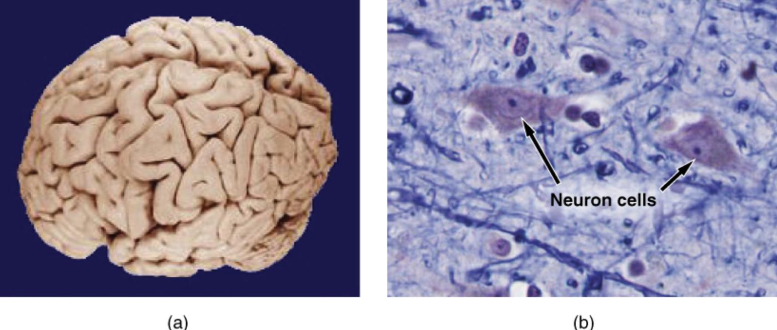
Neuron: The neuron is the basic functional unit of the nervous system, transmitting signals via electrical and chemical processes. Each neuron consists of a cell body, dendrites, and an axon, forming complex neural networks.
Dendrite: These branched extensions receive signals from other neurons, channeling them toward the cell body. Dendrites play a key role in synaptic communication and signal integration.
Axon: The axon is a long, slender projection that conducts electrical impulses away from the neuron’s cell body. It transmits signals to other neurons or target tissues, often over long distances.
Synapse: The synapse is the junction where neurons communicate, using neurotransmitters to relay signals. This structure is vital for memory formation and neural plasticity.
Gross Anatomy of the Human Brain
The brain’s macroscopic structure is a marvel of biological engineering. This section delves into the major regions visible in gross anatomy, focusing on their roles and significance.
- Cerebrum Overview: The cerebrum occupies most of the cranial cavity and is divided into frontal, parietal, temporal, and occipital lobes. Each lobe handles specialized functions, such as motor control, sensory perception, language, and vision.
- Cerebral Cortex Functionality: The cortex’s folded appearance increases its surface area, allowing for billions of neurons to process complex tasks. Damage to specific cortical areas can impair functions like speech or movement.
- Grey and White Matter Distribution: Grey matter forms the outer cortex and subcortical nuclei, while white matter lies beneath, forming tracts that connect distant brain regions. This organization ensures efficient communication across the brain.
- Functional Specialization: The cerebrum’s lobes work in concert, with the frontal lobe managing executive functions, the parietal lobe processing sensory data, the temporal lobe handling auditory and memory tasks, and the occipital lobe focusing on vision.
Microscopic Anatomy of the Nerve Cell
Neurons are the building blocks of the nervous system, with intricate structures enabling rapid communication. This section examines the microscopic features of neurons and their roles in neural networks.
- Neuron Structure: Each neuron consists of a cell body containing the nucleus, dendrites for receiving signals, and an axon for transmitting them. This polarized structure ensures directional signal flow.
- Dendritic Complexity: Dendrites branch extensively, increasing the surface area for synaptic connections. Their spines are dynamic, adapting to strengthen or weaken neural pathways during learning.
- Axon Functionality: Axons are insulated by myelin, produced by glial cells, which accelerates signal transmission. Nodes of Ranvier, gaps in the myelin sheath, further enhance conduction speed.
- Synaptic Transmission: Synapses use neurotransmitters like dopamine or glutamate to transmit signals. This chemical communication underpins processes like learning, memory, and motor control.
The Role of Grey and White Matter
Grey and white matter are integral to brain function, with distinct yet complementary roles. This section explores their contributions to neural processing and connectivity.
- Grey Matter’s Processing Power: Grey matter houses neuronal cell bodies and synapses, making it the site of information processing. Its density in the cortex supports complex cognitive tasks.
- White Matter’s Connectivity: White matter tracts, such as the corpus callosum, link the brain’s hemispheres and regions. This connectivity is crucial for coordinating tasks like language and spatial awareness.
- Balance of Structure and Function: The interplay between grey and white matter ensures the brain operates as a cohesive unit. Disruptions, such as in demyelinating diseases, can impair this balance.
Neural Networks and Cognitive Functions
Neural networks, formed by interconnected neurons, underpin the brain’s ability to process information and generate behavior. This section highlights how these networks support cognition and memory.
- Network Formation: Neurons form networks through synaptic connections, creating circuits that process sensory input and generate responses. These networks are dynamic, adapting through experience.
- Cognitive Processes: Neural networks in the cortex support higher-order functions like problem-solving and decision-making. The hippocampus, rich in neural connections, is critical for memory formation.
- Plasticity and Learning: Synaptic plasticity, the ability of synapses to strengthen or weaken, underlies learning and memory. Experiences shape these connections, enhancing cognitive flexibility.
- Pathology and Networks: Disruptions in neural networks, such as in neurodegenerative diseases, can impair cognition. Understanding these networks is key to developing treatments.
Histological Examination of Brain Tissue
Histology reveals the cellular details of brain tissue, providing insights into its structure and function. This section discusses the techniques and findings of histological studies.
- Staining Techniques: Histological stains, like hematoxylin and eosin, highlight neuronal structures and glial cells. These techniques reveal the organization of grey and white matter.
- Cellular Details: Microscopic examination shows the morphology of neurons, including dendritic spines and axonal projections. Glial cells, such as astrocytes, support neuronal function.
- Pathological Insights: Histology can identify abnormalities, such as neuronal loss in neurodegenerative conditions. These findings guide diagnosis and research.
- Research Applications: Histological studies inform our understanding of brain development and disease. Advanced imaging techniques complement histology for comprehensive analysis.
The human brain and its neurons are a testament to the complexity of biological systems. By examining the gross anatomy of the cerebrum and the microscopic structure of neurons, we gain a deeper appreciation for the mechanisms driving thought, memory, and behavior. The interplay of grey and white matter, along with the intricate neural networks, enables the brain to perform its myriad functions. Continued research into brain anatomy and histology promises to unlock further insights into neurology and human potential, paving the way for advancements in medical science.
Additional SEO Titles:
- Exploring the Human Brain: Gross and Microscopic Anatomy Unveiled
- Understanding Neurons: The Microscopic Structure of Nerve Cells
- Brain Anatomy 101: Cerebrum, Cortex, and Neural Networks Explained
- The Science of the Brain: Grey Matter, White Matter, and Synapses
- A Deep Dive into Brain and Neuron Anatomy for Neuroscience Enthusiasts

