Human Vertebrae Anatomy: A Comprehensive Guide to Spinal Structure

Date:

The human vertebral column is a marvel of biological engineering, serving as both protection for our nervous system and the central support structure for our body. Understanding its anatomy is crucial for healthcare professionals, students, and anyone interested in spine health. Let’s explore each component of a typical vertebra and its vital functions.

Human Vertebrae Anatomy: A Comprehensive Guide to Spinal Structure

Key Components of Vertebral Anatomy
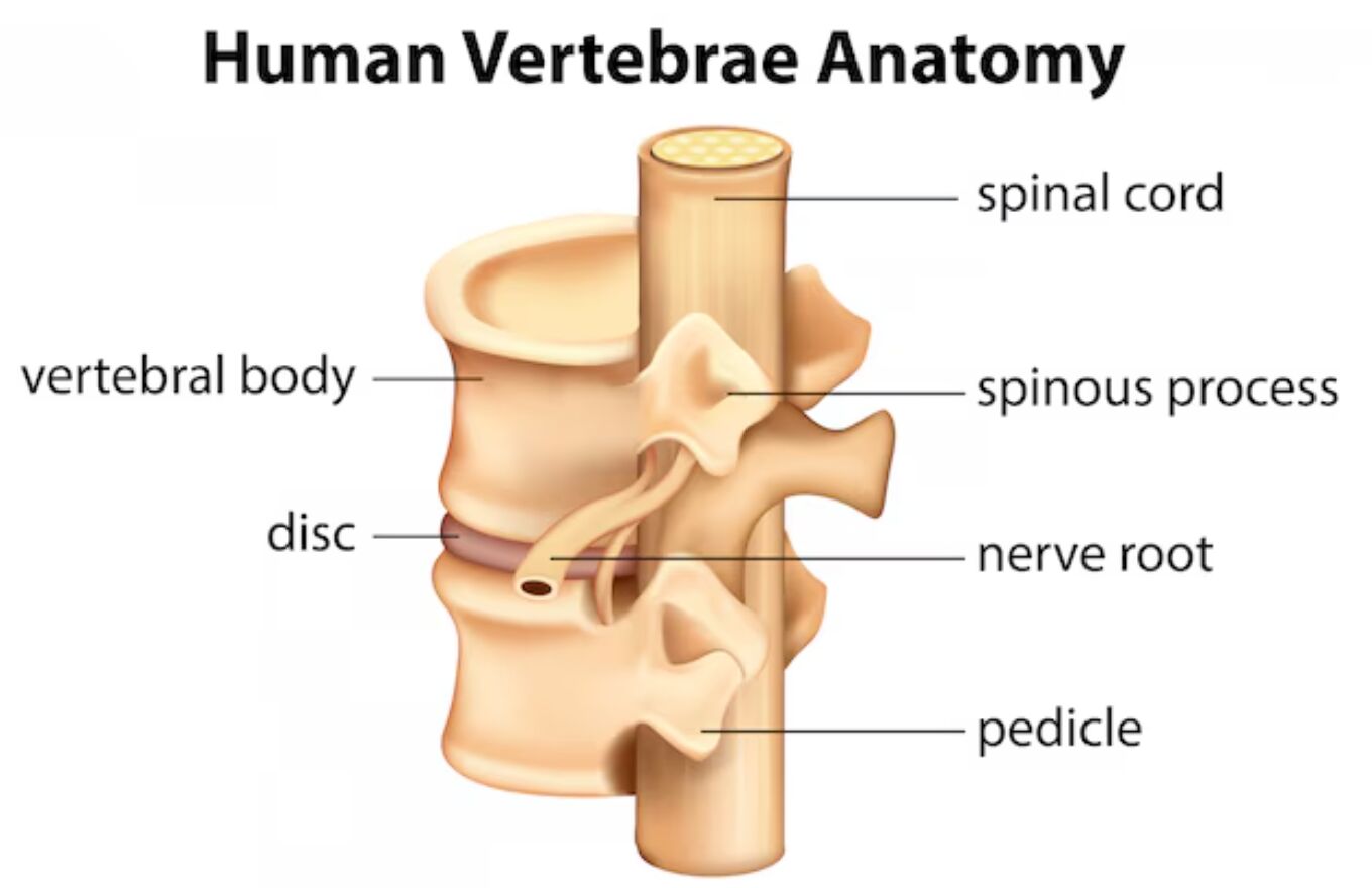
Vertebral Body

The vertebral body forms the main weight-bearing portion of the vertebra. This cylindrical structure of dense bone provides:
– Primary support for the spine’s weight-bearing function
– Attachment point for intervertebral discs
– Foundation for spinal stability and movement

Spinal Cord

Running through the central canal of the vertebrae, the spinal cord is crucial for:
– Transmitting nerve signals between brain and body
– Coordinating reflexes and movements
– Protected by the surrounding vertebral structures

- Advertisement -

Recommended Study Resource

Gray's Anatomy: The Anatomical Basis of Clinical Practice

Enhance your anatomical knowledge with Gray's Anatomy: The Anatomical Basis of Clinical Practice. This authoritative text offers in-depth insights and illustrations, perfect for medical students and practitioners aiming for clinical excellence.

Shop Now on Amazon

At AnatomyNote.com, we offer free resources on anatomy, pathology, and pediatric medicine for medical students and professionals. Purchasing through our Amazon links, like Gray's Anatomy, supports our server costs and content creation at no additional cost to you.

Disclosure: As an Amazon Associate, we earn a commission from qualifying purchases.

Disclosure: As an Amazon Associate, we earn a commission from qualifying purchases at no extra cost to you.

Spinous Process

This prominent posterior projection serves several purposes:
– Attachment point for muscles and ligaments
– Helps guide and limit spinal movement
– Provides leverage for spinal movements

Nerve Root

The nerve roots are vital components that:
– Branch off from the spinal cord
– Carry sensory and motor signals to specific body areas
– Exit through spaces between vertebrae

Pedicle

The pedicle is a crucial bridge-like structure that:
– Connects the vertebral body to posterior elements
– Provides pathway for blood vessels
– Forms part of the protective canal for nerve roots

Disc

The intervertebral disc serves as a crucial cushioning structure:
– Acts as a shock absorber between vertebrae
– Allows for spinal flexibility and movement
– Maintains proper spacing between vertebral bodies

- Advertisement -

Anatomy Flash Cards

Master anatomy with detailed, exam-ready flash cards.

Buy on Amazon

AnatomyNote.com offers free anatomy and pathology resources. Your purchase of Anatomy Flash Cards supports our site at no extra cost.

As an Amazon Associate, we earn from qualifying purchases.


Clinical Significance

Understanding vertebral anatomy is essential for:
– Diagnosing spinal conditions
– Planning surgical procedures
– Developing treatment strategies
– Understanding pain patterns
– Preventing spinal injuries


Common Conditions Affecting Vertebral Structures

Various conditions can affect different parts of the vertebrae:
– Herniated discs
– Spinal stenosis
– Vertebral fractures
– Nerve root compression
– Degenerative disc disease


Conclusion

The human vertebrae represent a complex and precisely engineered structure that provides both stability and flexibility to our spine while protecting the vital spinal cord. Each component plays a crucial role in maintaining proper spinal function and overall health. Understanding this anatomy is fundamental for healthcare professionals and helps patients better comprehend their spinal conditions and treatment options. As medical science advances, this knowledge continues to guide innovations in spine care and treatment approaches.

## Keywords:

We'd be thrilled to have your support!

Your generous contribution through a coffee keeps our passion alive.

Image source:

LEAVE A REPLY

Please enter your comment!
Please enter your name here

Share post:

Popular

spot_imgspot_img

Subscribe

More like this
Related

Vertebral Column Segments: A Comprehensive Anatomical Guide

The vertebral column represents one of the most remarkable anatomical structures in the human body, combining flexibility with structural support and neural protection. This complex system consists of distinct segments, each with unique characteristics that contribute to specific functional requirements. Understanding these segments is crucial for medical professionals in diagnosing and treating spinal conditions, as well as performing surgical interventions.

Vertebral Column: Anatomical Structure, Curvatures, and Functional Role

The vertebral column, a central pillar of the human body, comprises 24 vertebrae along with the sacrum and coccyx, segmented into cervical, thoracic, and lumbar regions with distinct curvatures. These regions—cervical (C1–C7), thoracic (T1–T12), and lumbar (L1–L5)—form primary (thoracic and sacrococcygeal) and secondary (cervical and lumbar) curves that support posture and movement. This article explores the anatomy, physical characteristics, and functional significance of the vertebral column, providing a comprehensive guide for understanding its role in human anatomy.

Cervical Spine Dynamics: A Comprehensive Analysis of Flexion-Extension X-ray Radiographs

Dynamic cervical spine imaging through flexion-extension radiographs provides crucial insights into the biomechanical function and stability of the cervical vertebrae. These specialized X-rays capture the spine's range of motion and help identify potential instabilities or abnormalities that might not be apparent in static imaging. Understanding the dynamic aspects of cervical spine mobility is essential for accurate diagnosis and treatment planning in various cervical pathologies.

Complete Guide to Vertebral Column Segments: From Cervical to Coccygeal Anatomy

The vertebral column represents the central supporting structure of the human body, comprising distinct segments each with unique anatomical and functional characteristics. This comprehensive color-coded illustration demonstrates the five major regions of the spine: cervical (red), thoracic (blue), lumbar (yellow), sacral (green), and coccygeal (purple). Understanding these segments is crucial for medical professionals in diagnosing and treating spinal conditions, as each region presents specific clinical challenges and considerations.