The sole of the foot is a marvel of muscular complexity, with layers of muscles working together to support weight and enable intricate movements. This article delves into the superficial, intermediate, and deep muscles of the left sole, presented through detailed plantar view diagrams, to provide a comprehensive examination of their anatomical structure and functional roles. These muscles, spanning three layers, are primarily responsible for flexing and extending the toes while providing the strength to counterbalance body weight, with each layer contributing uniquely to foot stability and locomotion. By analyzing the labeled illustrations, readers can gain a thorough understanding of these muscles’ significance in foot function and their relevance in clinical contexts.
Introduction to the Muscles of the Left Sole
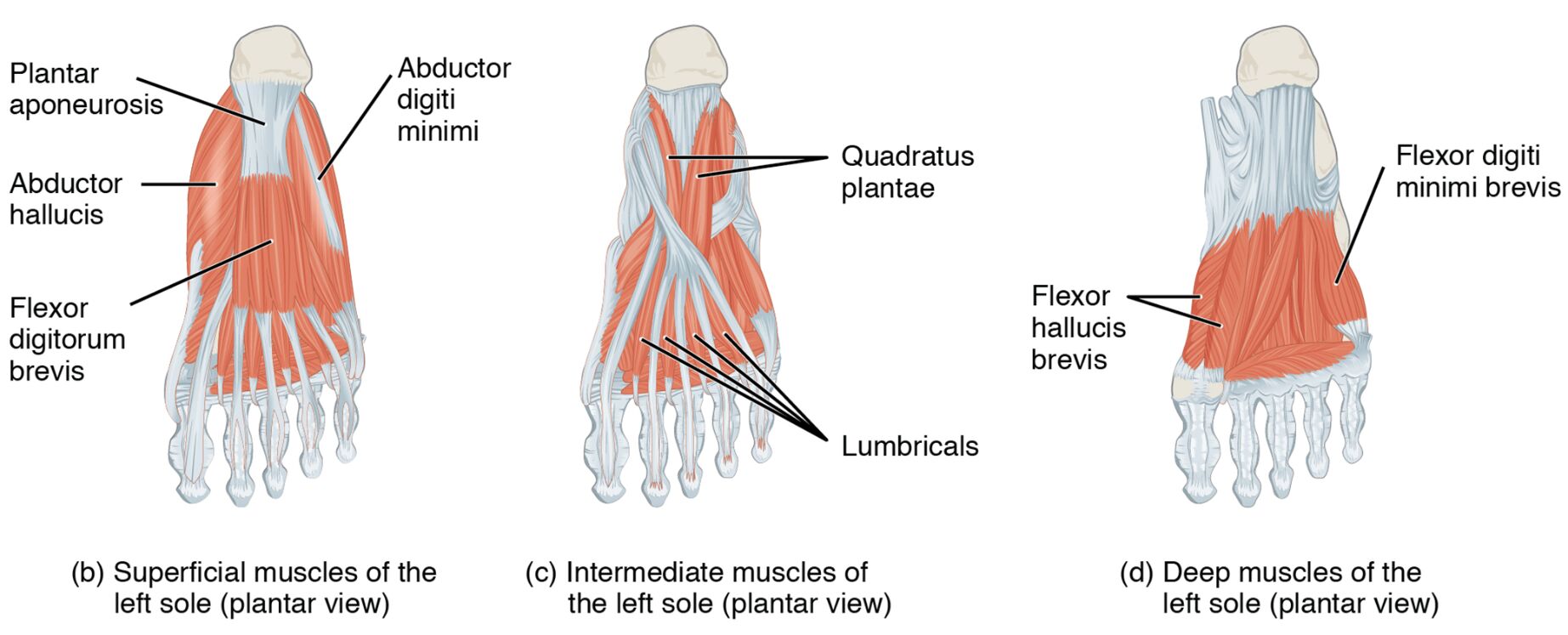
The superficial, intermediate, and deep muscles of the left sole form a multi-layered network across the plantar surface. Their plantar views highlight their diverse roles in supporting the foot. This section details the labeled structures that define their anatomy and function.
- Abductor hallucis: Positioned in the superficial layer along the medial edge, it abducts and flexes the big toe. It supports the medial arch and enhances foot balance.
- Flexor digitorum brevis: Located centrally in the superficial layer, it flexes the lesser toes. It provides grip strength and aids in weight distribution.
- Abductor digiti minimi: Found in the superficial layer along the lateral edge, it abducts and flexes the little toe. It contributes to lateral foot stability and movement.
- Lumbricals: Positioned in the intermediate layer centrally, these muscles flex the metatarsophalangeal joints and extend the interphalangeal joints. They enhance toe dexterity and assist in gripping the ground.
- Quadratus plantae (flexor accessorius): Located in the intermediate layer deeper, it assists in flexing the toes. It supports the flexor digitorum longus in efficient toe movement.
- Flexor hallucis brevis: Positioned in the deep layer near the big toe, it flexes the proximal phalanx of the big toe. It supports propulsion and maintains toe stability during push-off.
- Adductor hallucis (oblique head): Located in the deep layer medially, it adducts and flexes the big toe. It stabilizes the toe and reinforces the transverse arch.
- Adductor hallucis (transverse head): Found in the deep layer across the metatarsal heads, it adducts the big toe and supports the transverse arch. It aids in maintaining foot alignment under load.
- Flexor digiti minimi brevis: Positioned in the deep layer near the little toe, it flexes the proximal phalanx of the little toe. It enhances lateral toe movement and stability.
- Plantar interossei: Located in the deep layer between the metatarsals, these muscles adduct the toes toward the third toe. They assist in fine-tuning toe positioning and balance.
- Dorsal interossei: Positioned in the deep layer between the metatarsals, these muscles abduct the toes away from the third toe. They contribute to toe spreading and lateral stability.
The superficial, intermediate, and deep muscles of the left sole‘s layered structure provides comprehensive support. Their labeled views offer a detailed perspective on their anatomical and functional roles.
Functional Roles of the Sole Muscles
The superficial, intermediate, and deep muscles of the left sole are essential for diverse toe and foot movements. Their organization across layers enhances strength and flexibility. This section outlines their specific functional contributions.
- The abductor hallucis and flexor digitorum brevis in the superficial layer flex the big and lesser toes. They provide grip and support the medial arch during standing.
- The abductor digiti minimi in the superficial layer flexes and abducts the little toe. This action enhances lateral stability and balance.
- The lumbricals and quadratus plantae in the intermediate layer flex and extend the toes. They improve toe control and assist in weight distribution.
- The flexor hallucis brevis and adductor hallucis in the deep layer flex and adduct the big toe. They power propulsion and reinforce arch stability.
- The flexor digiti minimi brevis, plantar interossei, and dorsal interossei in the deep layer flex, adduct, and abduct the toes. These actions fine-tune toe positioning and lateral support.
The superficial, intermediate, and deep muscles of the left sole‘s coordinated efforts optimize foot performance. Their layered design ensures effective support and movement.
Clinical Significance and Practical Applications
The superficial, intermediate, and deep muscles of the left sole are frequently evaluated in clinical assessments of foot health. Their condition directly impacts mobility and stability. This section explores their clinical relevance.
- Weakness in the abductor hallucis can lead to flat feet or hallux valgus. Strengthening exercises help restore arch support and toe alignment.
- Strain in the flexor digitorum brevis may cause toe pain or claw toe deformity. Stretching and conditioning alleviate discomfort and improve function.
- Injury to the lumbricals can impair toe extension, affecting gait. Targeted therapy restores dexterity and coordination.
- Overuse of the adductor hallucis may contribute to bunion formation or metatarsalgia. Rest and rehabilitation prevent further strain.
- Understanding their anatomy aids in diagnosing conditions like plantar fasciitis. This knowledge guides effective treatment and preventive strategies.
This insight is valuable for professionals addressing foot concerns. The superficial, intermediate, and deep muscles of the left sole‘s roles underscore the need for precise therapeutic interventions.
Conclusion
The superficial, intermediate, and deep muscles of the left sole, as depicted in the plantar view, illustrate the foot’s intricate muscular framework that supports weight-bearing and movement across multiple layers. This article has explored their anatomical structure, diverse functional roles, and clinical significance, providing a thorough understanding of their importance. From the abductor hallucis stabilizing the medial arch to the dorsal interossei promoting toe spreading, each muscle contributes uniquely to foot stability and locomotion. Continued study of these muscles will enhance therapeutic approaches and deepen appreciation for the complex mechanics of the foot.

