The posterior aspect of the lower body houses a complex network of muscles that are essential for movement and stability. This article examines the pelvic and thigh muscles of the right leg, presented in a posterior view, to provide an in-depth look at their anatomical structure and functional significance. These powerful muscles, originating from the pelvic girdle and femur, play a crucial role in flexing the lower leg, extending the thigh, and facilitating adduction, abduction, and rotation of the thigh and lower leg. Through the labeled diagram, readers can gain a comprehensive understanding of these muscles’ contributions to leg function and their relevance in clinical practice.
Introduction to the Pelvic and Thigh Muscles
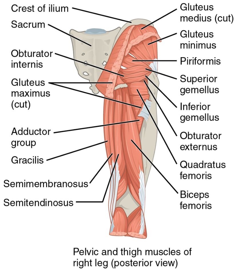
The pelvic and thigh muscles in the posterior view reveal a robust muscular framework. Their strategic placement supports a variety of lower limb movements. This section explores the labeled components that define their anatomy and roles.
- Crest of ilium: This bony ridge provides attachment for posterior hip muscles. It offers a stable base for movements involving the pelvis and thigh.
- Sacrum: This triangular bone anchors several pelvic muscles. It serves as a foundational support for hip and thigh stability.
- Obturator internus: A deep hip muscle, it laterally rotates the thigh. It enhances hip joint stability during rotational movements.
- Gluteus maximus (cut): The largest buttock muscle, it extends and laterally rotates the thigh. It provides significant power for standing and climbing.
- Adductor group: Comprising multiple muscles, this group adducts the thigh toward the midline. It supports balance and lateral movement control.
- Gracilis: Located on the inner thigh, this muscle adducts the thigh and flexes the knee. It aids in stabilizing the leg during rotation.
- Semimembranosus: A hamstring muscle, it flexes the knee and extends the thigh. It contributes to posterior thigh strength and stability.
- Semitendinosus: Another hamstring muscle, it flexes the knee and extends the thigh. It supports knee joint stability and leg flexion.
- Gluteus medius (cut): Positioned under the gluteus maximus, it abducts and medially rotates the thigh. It stabilizes the pelvis during walking.
- Gluteus minimus: The deepest gluteal muscle, it abducts and medially rotates the thigh. It assists in maintaining pelvic balance.
- Piriformis: Located deep in the buttock, it laterally rotates the thigh. It also stabilizes the hip joint during movement.
- Superior gemellus: A small muscle above the obturator internus, it laterally rotates the thigh. It supports fine-tuning of hip rotation.
- Inferior gemellus: Positioned below the obturator internus, it laterally rotates the thigh. It aids in stabilizing the hip joint.
- Obturator externus: A deep hip muscle, it laterally rotates the thigh. It enhances hip stability during complex movements.
- Quadratus femoris: A deep hip muscle, it laterally rotates and adducts the thigh. It provides additional support to the hip joint.
- Biceps femoris: Part of the hamstrings, it flexes the knee and extends the thigh. It is crucial for lower leg movement and stability.
The pelvic and thigh muscles‘ posterior arrangement ensures effective movement. Their detailed labeling provides insight into their structural and functional roles.
Functional Roles of the Pelvic and Thigh Muscles
The pelvic and thigh muscles are vital for a range of lower body actions. Their coordinated functions support strength, stability, and flexibility. This section details their specific contributions.
- The semimembranosus and semitendinosus flex the lower leg at the knee. They also extend the thigh, aiding in standing from a seated position.
- The gluteus maximus extends and laterally rotates the thigh. This action powers movements like running or climbing stairs.
- The adductor group adducts the thigh toward the midline. This movement is essential for maintaining balance during lateral steps.
- The gluteus medius and gluteus minimus abduct the thigh. They prevent the opposite side from dropping, ensuring pelvic stability during gait.
- The piriformis and obturator internus laterally rotate the thigh. These muscles are key for rotational actions like turning the leg outward.
- The biceps femoris supports knee flexion and thigh extension. Its dual role enhances lower leg mobility and strength.
The pelvic and thigh muscles‘ multifunctionality drives lower body performance. Their posterior positioning optimizes movement efficiency and stability.
Clinical Significance and Practical Applications
The pelvic and thigh muscles are often evaluated in clinical settings to assess leg function. Their health directly impacts mobility and quality of life. This section explores their clinical relevance.
- Strain in the semimembranosus or semitendinosus can cause hamstring injuries. Rehabilitation focuses on stretching and strengthening to restore flexibility.
- Weakness in the gluteus medius may lead to a Trendelenburg gait. Targeted exercises improve pelvic stability and walking ability.
- Injury to the gluteus maximus can impair thigh extension. Physical therapy helps regain power for daily activities like standing.
- Overuse of the adductor group may result in groin strain. Rest and conditioning alleviate pain and prevent further damage.
- Understanding their anatomy aids in diagnosing conditions like piriformis syndrome. This knowledge guides effective treatment and prevention strategies.
This insight is crucial for professionals addressing leg issues. The pelvic and thigh muscles‘ roles highlight the need for precise therapeutic interventions.
Conclusion
The pelvic and thigh muscles of the right leg, as depicted in the posterior view, illustrate the lower body’s intricate muscular design. This article has explored their anatomical structure, diverse functional roles, and clinical significance, providing a thorough understanding of their importance. From the gluteus maximus powering thigh extension to the semimembranosus supporting knee flexion, each muscle contributes uniquely to mobility and stability. Continued study of these muscles will enhance therapeutic approaches and deepen appreciation for the complex mechanics of leg movement.

