The deep musculature of the pelvic and thigh regions forms the foundation of the lower body’s strength and mobility. This article explores the deep pelvic and thigh muscles of the right leg, presented in an anterior view, offering a detailed look at their anatomical structure and functional roles. These muscles, originating from the pelvic girdle and inserting into the femur or knee joint, are vital for movements such as thigh flexion, lower leg extension, and a combination of adduction, abduction, and rotation. Through the labeled diagram, readers can develop a thorough understanding of these muscles’ contributions to leg function and their relevance in clinical settings.
Introduction to the Deep Pelvic and Thigh Muscles
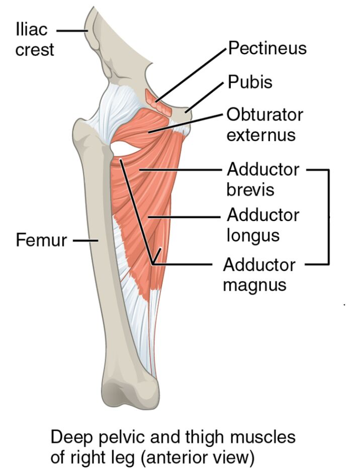
The deep pelvic and thigh muscles lie beneath the surface, providing critical support and movement. Their anterior view reveals a complex network essential for lower limb stability. This section details the labeled components that define their anatomy and purpose.
- Iliac crest: This bony ridge of the ilium serves as an attachment for deep hip muscles. It provides a stable anchor for movements involving the pelvis and thigh.
- Pectineus: Located near the pubic bone, this muscle flexes and adducts the thigh. It supports stability during medial leg motions and weight-bearing activities.
- Pubis: Part of the pelvic bone, it acts as an origin for adductor muscles. It facilitates thigh adduction and contributes to pelvic integrity.
- Obturator externus: A deep hip muscle, it laterally rotates the thigh. It enhances hip joint stability during rotational movements.
- Adductor brevis: Positioned above the adductor longus, this muscle adducts and flexes the thigh. It assists in maintaining thigh alignment during motion.
- Adductor longus: Found on the medial thigh, this muscle adducts the thigh toward the midline. It is crucial for balance and lateral movement control.
- Adductor magnus: A large medial thigh muscle, it adducts and extends the thigh. It delivers significant power for activities like standing or squatting.
- Femur: The thigh bone serves as an insertion point for these muscles. It provides the structural framework for lower limb movements and stability.
The deep pelvic and thigh muscles‘ deep placement ensures robust support. Their anterior perspective highlights their role in coordinating complex leg actions.
Functional Roles of the Deep Pelvic and Thigh Muscles
The deep pelvic and thigh muscles are essential for precise and powerful lower body movements. Their functions support a range of activities requiring strength and control. This section outlines their specific contributions to leg function.
- The pectineus and adductor brevis flex and adduct the thigh. These actions are vital for maintaining balance during lateral steps or pivoting.
- The adductor longus and adductor magnus adduct the thigh toward the midline. This movement enhances stability during walking or side-to-side shifts.
- The obturator externus laterally rotates the thigh. This function is key for rotational movements, such as turning the leg outward.
- The adductor magnus also extends the thigh, providing power for standing from a seated position. This dual role supports both strength and endurance.
- The femur acts as a lever for these muscles’ actions. Its alignment influences the efficiency of thigh and lower leg movements.
The deep pelvic and thigh muscles‘ coordinated efforts optimize leg performance. Their deep location ensures stability and precision in every motion.
Clinical Significance and Practical Applications
The deep pelvic and thigh muscles are frequently evaluated in clinical assessments of leg health. Their condition directly affects mobility and overall well-being. This section explores their clinical importance.
- Strain in the adductor longus can lead to groin pain or difficulty with adduction. Stretching and strengthening exercises help restore function and prevent recurrence.
- Injury to the obturator externus may impair thigh rotation. Physical therapy targeting this muscle can improve hip joint stability.
- Weakness in the adductor magnus can affect thigh extension and adduction. Rehabilitation focuses on rebuilding strength for daily activities.
- The pectineus‘ overactivity may contribute to hip flexor tightness. Targeted stretching alleviates discomfort and enhances flexibility.
- Understanding their anatomy aids in diagnosing conditions like hip adductor tendinopathy. This knowledge guides effective treatment plans and preventive measures.
This insight is invaluable for professionals addressing leg issues. The deep pelvic and thigh muscles‘ roles underscore the importance of targeted therapeutic interventions.
Conclusion
The deep pelvic and thigh muscles of the right leg, as illustrated in the anterior view, reveal the lower body’s intricate muscular support system. This article has examined their anatomical structure, diverse functional roles, and clinical significance, offering a comprehensive understanding of their importance. From the pectineus aiding thigh flexion to the adductor magnus powering extension, each muscle plays a unique role in leg mobility and stability. Continued exploration of these muscles will enhance therapeutic strategies and deepen appreciation for the sophisticated mechanics of lower limb movement.

