The hip and thigh region houses some of the body’s most powerful and essential muscles, critical for mobility and stability. This article explores the hip and thigh muscles through detailed anatomical diagrams of the right leg, showcasing both anterior and posterior views to highlight their structure and function. These muscles, originating from the pelvic girdle and femur, play a vital role in moving the femur, lower leg, and knee joint, with specific actions including flexion, extension, abduction, adduction, and rotation. By examining the labeled illustrations, readers can gain a thorough understanding of these muscles’ contributions to lower limb movement and their clinical relevance.
Introduction to the Hip and Thigh Muscles
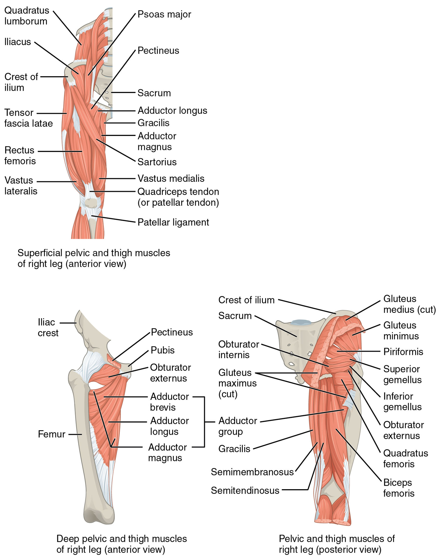
The hip and thigh muscles form a robust network that supports the lower body’s dynamic movements. Their strategic origins and insertions provide the foundation for a range of motions. This section reviews the labeled components that define their anatomical layout.
- Quadratus lumborum: Located near the lumbar region, this muscle assists in lateral flexion of the spine. It also stabilizes the pelvis during hip movements, enhancing overall posture.
- Iliacus: Positioned within the iliac fossa, this muscle works with the psoas major to flex the thigh. It plays a key role in lifting the leg during walking or climbing.
- Crest of ilium: This bony landmark serves as an attachment point for several hip muscles. It provides structural support and stability to the pelvic girdle.
- Tensor fasciae latae: Found on the lateral thigh, this muscle stabilizes the knee by tensing the iliotibial band. It also aids in thigh flexion and abduction.
- Rectus femoris: Part of the quadriceps group, this muscle extends the lower leg at the knee. It also contributes to thigh flexion, crucial for activities like kicking.
- Vastus lateralis: Another quadriceps muscle, located on the lateral thigh, it extends the knee. It supports powerful leg movements during running or jumping.
- Psoas major: Originating from the lumbar spine, this muscle flexes the hip joint. It works in tandem with the iliacus for thigh movement.
- Pectineus: Situated near the pubic bone, this muscle assists in thigh flexion and adduction. It supports stability during lateral leg movements.
- Sacrum: This triangular bone serves as an attachment site for pelvic muscles. It anchors the hip muscles, facilitating lower body stability.
- Adductor longus: Located on the medial thigh, this muscle adducts the thigh toward the midline. It is essential for maintaining balance during walking.
- Gracilis: Positioned along the inner thigh, this muscle adducts the thigh and flexes the knee. It aids in stabilizing the leg during rotation.
- Adductor magnus: A large medial thigh muscle, it adducts and extends the thigh. It provides significant power for movements like squatting.
- Sartorius: The longest muscle in the body, it flexes and rotates the thigh laterally. It also assists in knee flexion during crossing the legs.
- Vastus medialis: Part of the quadriceps, this muscle extends the knee and stabilizes the patella. It is critical for proper knee alignment.
- Quadriceps tendon (or patellar tendon): This strong tendon connects the quadriceps to the patella. It transmits force to extend the lower leg.
- Patellar ligament: Extending from the patella to the tibia, this ligament supports knee extension. It ensures efficient force transfer during movement.
- Iliac crest: This ridge of the ilium provides attachment for hip abductors. It contributes to pelvic stability and muscle leverage.
- Pubis: Part of the pelvic bone, it serves as an origin for adductor muscles. It supports thigh adduction and medial rotation.
- Obturator externus: A deep hip muscle, it laterally rotates the thigh. It stabilizes the hip joint during rotational movements.
- Adductor brevis: Located above the adductor longus, it adducts and flexes the thigh. It assists in maintaining thigh alignment.
- Obturator internus: A deep pelvic muscle, it laterally rotates the thigh. It supports hip joint stability during complex movements.
- Gluteus maximus (cut): The largest buttock muscle, it extends and laterally rotates the thigh. It provides power for standing and climbing.
- Gluteus medius (cut): Positioned under the gluteus maximus, it abducts and medially rotates the thigh. It stabilizes the pelvis during walking.
- Gluteus minimus: The deepest gluteal muscle, it abducts and rotates the thigh medially. It aids in maintaining pelvic balance.
- Piriformis: Located deep in the buttock, it laterally rotates the thigh. It also stabilizes the hip joint during movement.
- Superior gemellus: A small muscle above the obturator internus, it laterally rotates the thigh. It supports hip joint stability.
- Inferior gemellus: Positioned below the obturator internus, it also laterally rotates the thigh. It assists in fine-tuning hip rotation.
- Quadratus femoris: A deep hip muscle, it laterally rotates and adducts the thigh. It provides additional stability to the hip joint.
- Biceps femoris: Part of the hamstrings, it flexes the knee and extends the thigh. It is crucial for lower leg movement and stability.
- Semimembranosus: Another hamstring muscle, it flexes the knee and extends the thigh. It supports posterior thigh strength.
- Semitendinosus: Part of the hamstrings, it flexes the knee and extends the thigh. It aids in stabilizing the knee joint.
- Femur: The thigh bone serves as an insertion point for many muscles. It provides the structural foundation for lower limb movements.
The hip and thigh muscles‘ diverse origins and insertions enable complex motions. Their anatomical arrangement supports both strength and flexibility in the lower body.
Functional Roles of the Hip and Thigh Muscles
The hip and thigh muscles are essential for a wide range of movements. Their coordinated actions ensure efficient locomotion and stability. This section outlines their specific functions and contributions.
- The anterior muscles, like the rectus femoris and vastus lateralis, extend the lower leg at the knee. They also assist in thigh flexion, supporting activities like stepping up.
- The posterior muscles, including the biceps femoris and semimembranosus, flex the lower leg at the knee. They also extend the thigh, aiding in standing from a seated position.
- The adductor longus and adductor magnus adduct the thigh toward the midline. This action is vital for maintaining balance during lateral movements.
- The gluteus medius and gluteus minimus abduct the thigh, stabilizing the pelvis. They prevent the opposite side from dropping during walking.
- The piriformis and obturator internus rotate the thigh laterally. These muscles are key for rotational movements like turning the leg outward.
- The gluteus maximus extends the thigh, providing power for climbing or running. It also assists in lateral rotation for added versatility.
The hip and thigh muscles‘ multifunctionality enhances lower body performance. Their interplay ensures smooth and powerful movement across various activities.
Clinical Significance and Practical Applications
The hip and thigh muscles are critical in clinical assessments of lower limb function. Their health directly impacts mobility and quality of life. This section explores their clinical relevance.
- Injuries to the quadriceps tendon can impair knee extension, affecting walking ability. Rehabilitation focuses on strengthening these muscles to restore function.
- Strain in the hamstrings (e.g., biceps femoris) can limit knee flexion. Targeted exercises help recover flexibility and strength.
- Weakness in the gluteus medius may lead to Trendelenburg gait. Strengthening this muscle improves pelvic stability during movement.
- The adductor group‘s overactivity can cause groin strain. Stretching and conditioning alleviate discomfort and prevent injury.
- Understanding their anatomy aids in diagnosing conditions like hip osteoarthritis. This knowledge guides physical therapy and surgical planning.
This insight is valuable for professionals working with lower limb issues. The hip and thigh muscles‘ roles underscore the need for precise therapeutic interventions.
Conclusion
The hip and thigh muscles, as illustrated in the anterior and posterior views of the right leg, demonstrate the lower body’s remarkable anatomical complexity. This article has detailed their structure, diverse functional roles, and clinical importance, offering a comprehensive overview of their significance. From the rectus femoris extending the lower leg to the gluteus maximus powering thigh extension, each muscle contributes uniquely to mobility and stability. Continued exploration of these muscles will enhance therapeutic strategies and deepen appreciation for the intricate mechanics of human movement.

