The pelvic floor muscles are fundamental to supporting the pelvic organs, maintaining continence, and stabilizing the core against intra-abdominal pressure. This detailed exploration of the muscles of the pelvic floor in a superior view provides a comprehensive look at their anatomy and roles, offering valuable insights for understanding pelvic health.
Labeled Parts Introduction
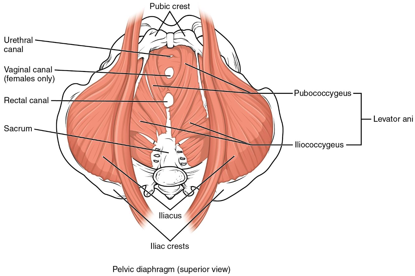
Pubic crest
The pubic crest is the superior border of the pubic bone, serving as an anterior attachment point for the pelvic floor muscles, and provides structural support to the pelvic girdle. It plays a key role in anchoring the muscles that support the bladder and other pelvic organs.
Urethral canal
The urethral canal is the passageway for urine from the bladder to the exterior, surrounded by pelvic floor muscles that act as a sphincter, and is essential for maintaining urinary continence. Its muscular support helps prevent involuntary leakage during physical activity.
Vaginal canal (females only)
The vaginal canal is the muscular canal in females that extends from the cervix to the vaginal opening, supported by the pelvic floor muscles, and is crucial for childbirth and sexual function. These muscles provide structural integrity and assist in controlling the canal’s tone.
Rectal canal
The rectal canal is the final segment of the large intestine, encircled by pelvic floor muscles that function as a sphincter, and is vital for fecal continence. Its muscular support helps regulate bowel movements and prevent leakage.
Sacrum
The sacrum is a triangular bone at the base of the spine, forming the posterior attachment for the pelvic floor muscles, and transmits weight from the upper body to the pelvis. It provides a stable foundation for the levator ani and other pelvic muscles.
Pubococcygeus
The pubococcygeus is a key muscle of the levator ani group, extending from the pubic bone to the coccyx, and supports the pelvic organs while aiding in sphincter control of the urethra and rectum. It is essential for maintaining pelvic floor strength and continence.
Levator ani
The levator ani is a broad muscle sheet forming the main component of the pelvic diaphragm, supporting the pelvic organs and resisting intra-abdominal pressure, and plays a critical role in maintaining continence. It works collectively with other muscles to stabilize the pelvic floor.
Iliococcygeus
The iliococcygeus is a posterior part of the levator ani, stretching from the ilium to the coccyx, and supports the pelvic floor by elevating the anus and aiding in continence. It contributes to the structural integrity of the pelvic diaphragm.
Iliacus
The iliacus is a muscle originating from the iliac fossa, extending to the femur, and indirectly supports the pelvic floor by stabilizing the pelvis. It works with the pelvic muscles to facilitate hip flexion and maintain pelvic alignment.
Iliac crests
The iliac crests are the superior borders of the ilium, providing lateral attachment points for the pelvic muscles, and contribute to the stability of the pelvic girdle. They support the overall structure of the pelvic floor and its muscular attachments.
Overview of Pelvic Floor Muscle Anatomy
The muscles of the pelvic floor form a hammock-like structure that supports the urethral canal, vaginal canal, and rectal canal, while resisting intra-abdominal pressure. This superior view highlights the levator ani, pubococcygeus, and iliococcygeus, which are crucial for maintaining organ position and continence. Their intricate arrangement underscores their importance in pelvic stability and function.
- Serves as a dynamic support system for organs like the bladder and rectum.
- Enhances core strength by counteracting pressure during activities like coughing.
Structure of Pelvic Floor Muscles
The levator ani, including the pubococcygeus and iliococcygeus, forms the primary muscular layer of the pelvic diaphragm, attaching to the pubic crest anteriorly and sacrum posteriorly. The iliacus and iliac crests provide additional support by stabilizing the pelvis, while the urethral canal and rectal canal are enveloped by sphincter-like muscles.
- The pubococcygeus is a central component of the levator ani, offering robust support.
- The iliococcygeus reinforces the posterior pelvic floor structure.
Role in Continence and Support
The levator ani and pubococcygeus act as sphincters around the urethral canal and rectal canal, preventing involuntary leakage by contracting during increased abdominal pressure. The vaginal canal in females is supported by these muscles, aiding in childbirth and maintaining tone, while the sacrum provides a stable anchor.
- The urethral canal relies on pelvic floor strength to maintain urinary control.
- The rectal canal benefits from muscular support during bowel movements.
Clinical Relevance and Physical Health
Weakness in the levator ani can lead to pelvic organ prolapse or incontinence, conditions often assessed by examining the pubococcygeus and iliococcygeus. The iliacus may be evaluated for pelvic tilt, which can affect pelvic floor function, requiring targeted exercises to restore balance.
- A weakened pubococcygeus may contribute to stress urinary incontinence.
- The iliac crests serve as landmarks for assessing pelvic alignment.
Physical Examination and Rehabilitation
Physical exams involve palpating the sacrum and pubic crest to assess pelvic floor muscle tone, while the iliacus is checked for hip flexibility. Rehabilitation programs often include Kegel exercises to strengthen the levator ani and pubococcygeus, improving continence and pelvic support.
- Proper tone in the levator ani enhances pelvic organ support.
- The iliococcygeus is targeted to address posterior pelvic floor weakness.
Conclusion
The muscles of the pelvic floor, as depicted in this superior view, create a resilient network that supports the urethral canal, vaginal canal, and rectal canal while maintaining core stability. Anchored by the sacrum, pubic crest, and iliac crests, with contributions from the iliacus, these muscles are vital for continence and physical health. Understanding their anatomy provides a foundation for addressing pelvic floor disorders and optimizing musculoskeletal function.

