Cell division is a critical process that ensures the accurate distribution of genetic material into two new nuclei, followed by the division of the cytoplasm to form two daughter cells. This article explores a detailed chart of the stages of mitosis and cytokinesis, providing a comprehensive view of each phase from prophase to cytokinesis, supported by microscopic images. By examining these stages, we gain insight into the mechanisms that drive growth, repair, and reproduction in eukaryotic cells.
Introduction to the Labeled Components
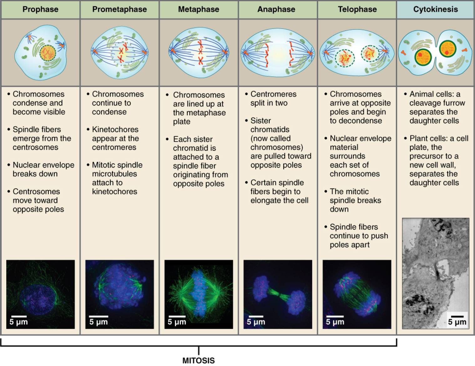
The chart includes six labeled stages of cell division, each representing a distinct phase with specific cellular events. Below is a detailed explanation of each labeled component, highlighting their roles in the process of mitosis and cytokinesis.
Prophase
Prophase marks the beginning of mitosis, where chromosomes condense and become visible, and the nuclear envelope starts to break down. Centrosomes move toward opposite poles, initiating the formation of the mitotic spindle fibers that will guide chromosome separation.
Prometaphase
Prometaphase follows prophase, with chromosomes continuing to condense as the nuclear envelope fully breaks down, allowing spindle fibers to attach to kinetochores. The mitotic spindle microtubules begin attaching to kinetochores, preparing chromosomes for alignment.
Metaphase
Metaphase is characterized by chromosomes aligning at the metaphase plate, a plane equidistant from the two spindle poles, ensuring equal distribution. Each chromosome is fully condensed, and spindle fibers originating from opposite poles are attached to its kinetochores.
Anaphase
Anaphase involves the centromeres splitting, separating sister chromatids (now called chromosomes) that are pulled toward opposite poles by spindle fibers. Certain spindle fibers elongate the cell, facilitating the physical separation of the genetic material.
Telophase
Telophase sees chromosomes arriving at opposite poles and beginning to decondense, with the nuclear envelope reforming around each set of chromosomes. The mitotic spindle breaks down, and spindle fibers continue to push the poles apart, marking the end of mitosis.
Cytokinesis
Cytokinesis is the final stage where the cytoplasm divides, separating the two daughter cells, with animal cells forming a cleavage furrow and plant cells developing a cell plate precursor to a new cell wall. This process completes cell division, resulting in two identical daughter cells.
Anatomical Overview of Cell Division
Cell division encompasses a series of orchestrated stages that ensure the faithful replication and distribution of genetic material. This section explores the anatomical features and progression through mitosis and cytokinesis as depicted in the chart.
- Chromosome Condensation: In prophase, chromatin condenses into discrete chromosomes, becoming visible under a microscope due to the coiling of DNA with histone proteins. This condensation is essential for their movement during mitosis.
- Spindle Apparatus Formation: During prometaphase and metaphase, the mitotic spindle, composed of microtubules, forms and organizes, with centrosomes acting as organizing centers. This apparatus is critical for aligning and separating chromosomes.
- Chromosome Alignment: Metaphase ensures chromosomes are positioned at the metaphase plate, a crucial checkpoint where the spindle attachment is verified, preventing aneuploidy. This alignment sets the stage for accurate division.
- Chromatid Separation: Anaphase drives the physical separation of sister chromatids, pulled by kinetochore microtubules shortening, while polar microtubules elongate the cell. This movement ensures each daughter cell receives a complete genome.
- Nuclear Reformation: In telophase, the reassembly of the nuclear envelope around decondensing chromosomes restores the nucleus, preparing the cell for interphase. This reformation signals the completion of nuclear division.
- Cytoplasmic Division: Cytokinesis varies between animal and plant cells, with animal cells using a contractile ring of actin and myosin to form a cleavage furrow, while plant cells deposit a cell plate. This division ensures each daughter cell has its own cytoplasm and organelles.
Physical Characteristics of Cell Division Stages
The physical changes during cell division stages are observable through microscopic imaging, reflecting the dynamic nature of cellular processes. This section examines these characteristics as shown in the chart’s micrographs.
- Prophase Appearance: Chromosomes appear as thick, thread-like structures, and the nuclear envelope begins to disintegrate, with centrosomes migrating to opposite ends. The cell’s outline remains intact, with a scale of 5 micrometers visible.
- Prometaphase Morphology: The nuclear envelope is absent, and chromosomes show partial alignment with spindle fibers attached, creating a chaotic yet organized appearance. The scale remains at 5 micrometers, highlighting cellular detail.
- Metaphase Structure: Chromosomes align in a straight line at the metaphase plate, with spindle fibers forming a symmetrical pattern, visible at 5 micrometers scale. This alignment is a critical visual checkpoint.
- Anaphase Configuration: Sister chromatids separate, moving toward opposite poles, with spindle fibers elongating the cell, observable at 5 micrometers scale. The cell elongates, indicating active division.
- Telophase Shape: The cell begins to assume two distinct nuclear regions, with chromosomes decondensing and the spindle breaking down, still at 5 micrometers scale. The nuclear envelope’s reformation is a key feature.
- Cytokinesis Form: In animal cells, a cleavage furrow indents the cell membrane, while in plant cells, a cell plate forms, both visible at 5 micrometers scale. This stage completes the physical separation of daughter cells.
Functional Significance of Cell Division
Cell division is vital for growth, repair, and reproduction, ensuring the continuity of genetic information. This section highlights the functional roles of each stage in cellular biology.
- Genetic Material Distribution: Prophase initiates chromosome condensation, setting the stage for their accurate segregation during mitosis, ensuring each daughter cell receives identical DNA. This process is fundamental for cell replication.
- Spindle Functionality: Prometaphase and metaphase establish the spindle apparatus, which aligns and attaches to chromosomes, facilitating their movement. This ensures precise genetic distribution during division.
- Chromosome Segregation: Anaphase separates sister chromatids, driven by microtubule dynamics, preventing genetic imbalances. This stage is critical for maintaining genomic stability.
- Nuclear Restoration: Telophase reforms the nuclear envelope, allowing the cell to resume interphase functions like transcription and protein synthesis. This restoration supports cellular homeostasis.
- Cytoplasmic Allocation: Cytokinesis divides the cytoplasm, ensuring each daughter cell inherits organelles and nutrients, supporting their independent survival. This allocation is essential for tissue development.
Implications for Cellular Health and Research
Cell division has significant implications for cellular health and scientific research, particularly in understanding diseases related to uncontrolled division. This section explores its broader impact and potential applications.
- Cancer Research: Abnormalities in mitosis, such as failure of the spindle checkpoint, can lead to aneuploidy, a common feature in cancer cells. Studying these stages informs the development of mitotic inhibitors like taxanes.
- Genetic Disorders: Errors in chromosome segregation during anaphase can cause conditions like Down syndrome, resulting from an extra chromosome 21. Research into mitotic fidelity helps prevent such anomalies.
- Therapeutic Strategies: Drugs targeting microtubules, such as vinblastine, disrupt mitosis in rapidly dividing cancer cells, offering a treatment avenue. This approach leverages the specificity of mitotic phases.
- Regenerative Medicine: Understanding cytokinesis supports the development of tissue engineering techniques, where controlled cell division is used to grow tissues. This application enhances regenerative therapies.
- Cell Cycle Regulation: Dysregulation of the mitotic cycle is linked to aging and senescence, as cells lose division capacity. Investigating these stages provides insights into extending cellular lifespan.
Cell division, through the precise stages of mitosis and cytokinesis, ensures the accurate replication and distribution of genetic material, underpinning growth and repair across organisms. Its intricate mechanisms and critical roles make it a focal point for advancing cellular biology and developing innovative medical treatments.

