The left humerus in its posterior view reveals critical anatomical landmarks and muscle attachment sites, essential for understanding upper limb function. This article provides a detailed exploration of the left humerus from the posterior perspective, offering valuable insights for medical students studying shoulder and elbow mechanics.
Labeled Anatomical Features
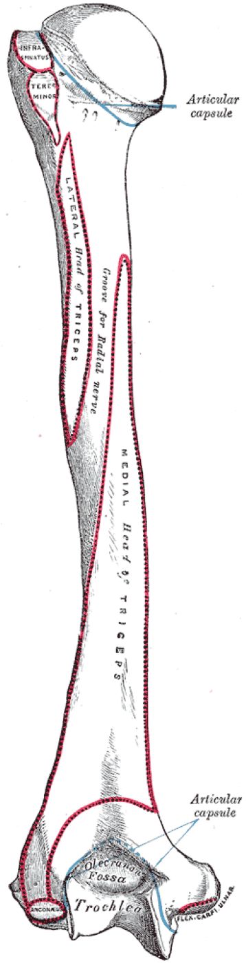
Articular Capsule
The articular capsule surrounds the shoulder joint and elbow joint, providing stability while allowing mobility. It attaches to the anatomical neck of the humerus proximally and the distal humerus at the elbow, encapsulating the joint spaces.
Infraspinatus
The infraspinatus muscle inserts onto the greater tubercle of the humerus, playing a key role in external rotation of the shoulder. It originates from the infraspinous fossa of the scapula, contributing to the stability of the shoulder joint as part of the rotator cuff.
Teres Minor
The teres minor inserts on the greater tubercle, just below the infraspinatus, assisting in external rotation and adduction of the arm. This rotator cuff muscle originates from the lateral border of the scapula, working alongside the infraspinatus to stabilize the shoulder.
Groove for Radial Nerve
The groove for the radial nerve, also known as the radial groove, is a shallow channel on the posterior humeral shaft where the radial nerve and profunda brachii artery travel. This groove protects the nerve as it wraps around the humerus, supplying the posterior arm and forearm muscles.
Head of Triceps
The head of the triceps, specifically the lateral and medial heads, originates from the posterior surface of the humeral shaft. These heads contribute to elbow extension, with the triceps being the primary extensor muscle of the forearm.
Olecranon Fossa
The olecranon fossa is a deep depression on the posterior distal humerus that accommodates the olecranon process of the ulna during elbow extension. This fossa ensures full extension of the elbow joint without impingement.
Trochlea
The trochlea is a spool-shaped structure on the distal humerus that articulates with the ulna, forming part of the elbow joint. It facilitates hinge-like flexion and extension of the forearm, ensuring stable movement.
Flexor Carpi Ulnaris
The flexor carpi ulnaris originates from the medial epicondyle of the humerus, contributing to wrist flexion and ulnar deviation. This muscle also has a humeral head, making it a key flexor of the forearm.
Detailed Anatomy of the Left Humerus: Posterior View
Overview of the Humerus
The humerus is the long bone of the upper arm, connecting the shoulder to the elbow through its complex structure. The posterior view highlights features critical for shoulder and elbow function, making it a key area of study for medical students.
- The humerus consists of a proximal end, shaft, and distal end, each with distinct anatomical landmarks.
- Its posterior surface is a primary attachment site for muscles that extend the elbow and externally rotate the shoulder.
- The bone articulates with the scapula at the shoulder joint and with the radius and ulna at the elbow joint.
- Its structure supports a wide range of upper limb movements, from lifting to throwing.
Proximal Humerus and Rotator Cuff Attachments
The proximal humerus in the posterior view showcases the greater tubercle and its rotator cuff muscle attachments. These structures are essential for shoulder stability and movement.
- The greater tubercle serves as the insertion site for the infraspinatus and teres minor, both rotator cuff muscles.
- The infraspinatus occupies the upper facet of the greater tubercle, facilitating external rotation of the arm.
- The teres minor inserts just below, contributing to shoulder stability during rotation.
- The anatomical neck, where the articular capsule attaches, marks the boundary of the humeral head’s articular surface.
Humeral Shaft and Radial Nerve Groove
The humeral shaft in the posterior view features the radial groove, a critical landmark for neurovascular structures. This area is significant for understanding nerve protection and muscle attachments.
- The radial groove runs diagonally across the posterior shaft, housing the radial nerve and profunda brachii artery.
- The radial nerve innervates the posterior arm and forearm muscles, making this groove clinically relevant for nerve injuries.
- The triceps brachii’s lateral and medial heads originate from the posterior shaft, above and below the groove.
- The shaft’s robust structure, composed of cortical bone, supports the mechanical stresses of arm movement.
Distal Humerus and Elbow Joint Features
The distal humerus in the posterior view includes the olecranon fossa and trochlea, which are integral to elbow joint function. These features ensure smooth forearm motion and stability.
- The olecranon fossa accommodates the ulna’s olecranon process during elbow extension, preventing overextension.
- The trochlea articulates with the ulna, forming a stable hinge joint for flexion and extension.
- The medial epicondyle, visible from the posterior view, serves as the origin for the flexor carpi ulnaris.
- The distal humerus is reinforced with cortical bone, providing strength to withstand the forces of elbow movement.
Functional Role of the Humerus in Movement
The humerus facilitates a wide range of movements through its muscle attachments and joint articulations, as seen in the posterior view. This functionality is crucial for daily activities and clinical assessment.
- The infraspinatus and teres minor, attaching to the greater tubercle, enable external rotation of the shoulder, essential for actions like throwing.
- The triceps brachii, originating from the posterior shaft, is the primary elbow extensor, critical for pushing movements.
- The radial groove protects the radial nerve, ensuring innervation to forearm extensors for wrist and finger extension.
- The trochlea and olecranon fossa work together to allow smooth elbow extension, supporting activities like reaching.
Clinical Relevance of the Humerus Posterior View
The posterior humerus is clinically significant due to its association with nerve injuries, fractures, and muscle pathologies. This knowledge is essential for diagnosing and treating related conditions.
- Radial nerve injuries in the radial groove can occur due to humeral shaft fractures, leading to wrist drop and impaired forearm extension.
- Distal humerus fractures involving the olecranon fossa can disrupt elbow extension, requiring surgical fixation.
- Rotator cuff injuries, such as tears to the infraspinatus or teres minor, may affect shoulder rotation and stability.
- Understanding the posterior humerus aids in surgical planning, such as for triceps repair or radial nerve decompression.
The left humerus in its posterior view provides a critical perspective on the bone’s role in upper limb function, from shoulder rotation to elbow extension. For medical students, mastering this anatomy enhances their ability to diagnose and manage upper limb conditions, deepening their understanding of musculoskeletal health.

