The cross-sectional anatomy of the neck at the sixth cervical vertebra level reveals the complex relationships between vital structures essential for head and neck function. This region demonstrates intricate layering of muscles, nerves, vessels, and visceral structures, making it crucial for medical professionals to understand these relationships for clinical practice and surgical intervention.
Labeled Anatomical Structures:
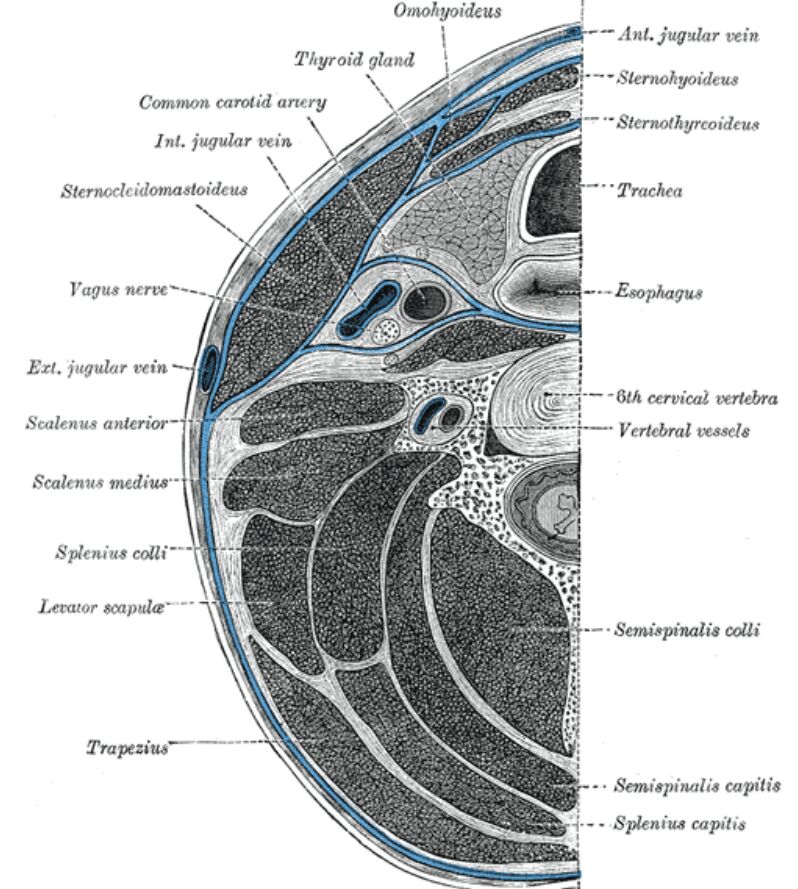
Omohyoideus: An infrahyoid muscle with superior and inferior bellies. This muscle depresses the hyoid bone and larynx while providing indirect tension to the deep cervical fascia.
Thyroid gland: An endocrine organ producing thyroid hormones (T3, T4) and calcitonin. The lateral lobes at this level contain rich vascular supply and are surrounded by a fibrous capsule.
Common carotid artery: The main arterial supply to the head and neck region. This vessel bifurcates into internal and external carotid arteries at the upper border of the thyroid cartilage.
Internal jugular vein: The principal venous drainage of the brain and superficial regions of the face and neck. This large vessel runs within the carotid sheath alongside the common carotid artery.
Sternocleidomastoideus: A prominent neck muscle responsible for head rotation and flexion. It serves as a key landmark for identifying underlying neurovascular structures.
Vagus nerve: The tenth cranial nerve carrying parasympathetic fibers to thoracic and abdominal viscera. It travels within the carotid sheath throughout the neck.
External jugular vein: A superficial vein draining the exterior of the head and neck. It crosses superficially to the sternocleidomastoid muscle before piercing the deep fascia.
Scalenus anterior: The most anterior of the scalene muscles, inserting into the first rib. This muscle serves as a crucial landmark for identifying the brachial plexus and subclavian vessels.
Scalenus medius: The middle scalene muscle attaching to the first rib posterior to the subclavian groove. This muscle, along with scalenus anterior, forms a potential space for neurovascular structures.
Splenius colli: A neck extensor muscle contributing to head and neck rotation. It works synergistically with other posterior neck muscles to control head position.
Levator scapulae: A muscle originating from the upper cervical vertebrae and inserting into the superior angle of the scapula. It functions to elevate and rotate the scapula.
Trapezius: A large superficial muscle of the posterior neck and upper back. It plays a crucial role in scapular positioning and head extension.
Semispinalis colli: A deep posterior neck muscle essential for spinal extension and rotation. It consists of multiple fascicles spanning several vertebral levels.
Semispinalis capitis: A thick muscle extending from the thoracic vertebrae to the occipital bone. It functions as a powerful head extensor.
Splenius capitis: A broad strap-like muscle in the back of the neck. It extends, laterally flexes, and rotates the head.
Vertebral vessels: The vertebral artery and accompanying veins passing through the transverse foramina. These vessels supply crucial circulation to the brain and upper spinal cord.
Anatomical Organization and Function
The cervical region at C6 demonstrates precise compartmentalization of structures. This level represents a crucial area where multiple vital structures converge, making it particularly important for surgical approaches and clinical assessment.
Fascial Relationships
The deep cervical fascia divides the neck into distinct compartments. This fascial organization provides pathways for structure organization while potentially limiting infection spread.
Clinical Significance
Diagnostic Applications
Understanding cross-sectional anatomy is essential for interpreting imaging studies. The relationships between structures help identify pathological conditions and guide intervention planning.
Surgical Considerations
Knowledge of these anatomical relationships is crucial for surgical approaches. Safe surgical corridors must be identified while protecting vital neurovascular structures.
- Cervical Cross-Sectional Anatomy: A Comprehensive Guide at C6 Level
- Understanding Neck Anatomy: Detailed Analysis at Sixth Cervical Vertebra
- Cross-Sectional Guide to Cervical Neurovascular and Muscular Structures
- C6 Level Neck Anatomy: Clinical and Surgical Considerations
- Cervical Anatomy in Cross-Section: Essential Guide for Medical Professionals

