The lacrimal bone, the smallest and most fragile of the facial bones, plays a crucial role in the formation of the orbit and lacrimal drainage system. This thin, scale-like bone contributes to the medial orbital wall and helps form the nasolacrimal duct, making it essential for proper tear drainage and orbital integrity. Understanding its anatomy is crucial for ophthalmologists, orbital surgeons, and medical professionals dealing with orbital pathologies.
By Henry Vandyke Carter – Henry Gray (1918) Anatomy of the Human Body (See "Book" section below)Bartleby.com: Gray's Anatomy, Plate 163, Public Domain, https://commons.wikimedia.org/w/index.php?curid=791891
Labeled Parts Introduction
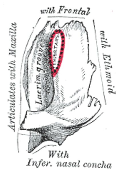
Articulation with Frontal The superior border that connects with the orbital plate of the frontal bone. This junction forms part of the superior orbital rim and provides stability to the medial orbital wall.
Articulation with Maxilla The anterior connection that meets the frontal process of the maxilla. This articulation helps form the lateral wall of the nasal cavity and contributes to the nasolacrimal canal.
Articulation with Inferior nasal concha The inferior portion that meets the inferior nasal concha. This junction contributes to the formation of the lateral nasal wall and nasolacrimal duct system.
Lacrimal Fossa A deep groove on the lateral surface (highlighted in red in the image). This fossa houses the lacrimal sac and forms the beginning of the nasolacrimal duct system.
Main Article Content
Anatomical Structure and Development
The lacrimal bone begins its development in the early stages of facial formation. This delicate structure ossifies from a single center appearing around the 12th week of fetal life.
The bone’s development is closely tied to the formation of the nasolacrimal system and orbital cavity. Its strategic position and relationships make it crucial for proper tear drainage and orbital function.
Structural Components
Orbital Surface
The orbital surface represents the primary functional component of the lacrimal bone. This surface contributes to the medial orbital wall and contains significant features:
- Smooth orbital surface
- Posterior lacrimal crest
- Lacrimal groove
- Attachment areas for orbital soft tissues
Nasal Surface
The nasal aspect demonstrates important relationships with the nasal cavity. This surface includes:
- Mucosal covering
- Contribution to middle meatus
- Relationship with ethmoid air cells
Clinical Significance
Surgical Considerations
The lacrimal bone’s location and fragility make it particularly important in various surgical procedures. Key considerations include:
- Dacryocystorhinostomy (DCR) Surgery:
- Access to nasolacrimal system
- Bone removal techniques
- Preservation of surrounding structures
- Orbital Surgery:
- Approach to medial orbit
- Protection of lacrimal drainage system
- Management of fractures
Pathological Conditions
- Nasolacrimal Duct Obstruction:
- Congenital versus acquired
- Impact on tear drainage
- Treatment approaches
- Orbital Fractures:
- Patterns involving lacrimal bone
- Associated injuries
- Management strategies
Radiological Assessment
Imaging the lacrimal bone requires specific techniques and understanding:
- CT for bony detail
- MRI for soft tissue evaluation
- Special views for nasolacrimal system
Conclusion
Understanding lacrimal bone anatomy is essential for medical professionals involved in orbital and lacrimal surgery. Its role in tear drainage and orbital structure makes it a critical component of facial anatomy. Advances in surgical techniques and imaging continue to emphasize the importance of detailed anatomical knowledge for optimal patient care.
- “Lacrimal Bone Anatomy: Essential Guide for Medical Professionals”
- “Understanding the Lacrimal Bone: Structure and Clinical Applications”
- “Comprehensive Analysis of Lacrimal Bone Anatomy”
- “Lacrimal Bone: From Basic Anatomy to Surgical Considerations”
- “Medical Guide to Lacrimal Bone Anatomy and Pathology”

